6MGQ

ERAP1 in the open conformation bound to 10mer phosphinic inhibitor DG014


Experimental Data Snapshot

  • Method: X-RAY DIFFRACTION
  • Resolution: 2.92 Å
  • R-Value Free: 0.256 
  • R-Value Work: 0.198 
  • R-Value Observed: 0.201 

wwPDB Validation   3D Report Full Report

Currently 6MGQ does not have a validation slider image.


This is version 3.2 of the entry. See complete history


Literature

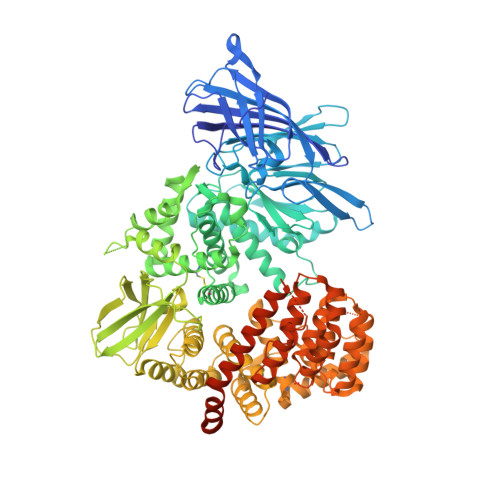
Conformational dynamics linked to domain closure and substrate binding explain the ERAP1 allosteric regulation mechanism.

Maben, Z.Arya, R.Georgiadis, D.Stratikos, E.Stern, L.J.

(2021) Nat Commun 12: 5302-5302

  • DOI: https://doi.org/10.1038/s41467-021-25564-w
  • Primary Citation of Related Structures:  
    6M8P, 6MGQ

  • PubMed Abstract: 

    The endoplasmic-reticulum aminopeptidase ERAP1 processes antigenic peptides for loading on MHC-I proteins and recognition by CD8 T cells as they survey the body for infection and malignancy. Crystal structures have revealed ERAP1 in either open or closed conformations, but whether these occur in solution and are involved in catalysis is not clear. Here, we assess ERAP1 conformational states in solution in the presence of substrates, allosteric activators, and inhibitors by small-angle X-ray scattering. We also characterize changes in protein conformation by X-ray crystallography, and we localize alternate C-terminal binding sites by chemical crosslinking. Structural and enzymatic data suggest that the structural reconfigurations of ERAP1 active site are physically linked to domain closure and are promoted by binding of long peptide substrates. These results clarify steps required for ERAP1 catalysis, demonstrate the importance of conformational dynamics within the catalytic cycle, and provide a mechanism for the observed allosteric regulation and Lys/Arg528 polymorphism disease association.


  • Organizational Affiliation

    Department of Pathology, University of Massachusetts Medical School, Worcester, MA, USA.


Macromolecules
Find similar proteins by:  (by identity cutoff)  |  3D Structure
Entity ID: 1
MoleculeChains Sequence LengthOrganismDetailsImage
Endoplasmic reticulum aminopeptidase 1
A, B, C
948Homo sapiensMutation(s): 0 
Gene Names: ERAP1APPILSARTS1KIAA0525UNQ584/PRO1154
EC: 3.4.11
UniProt & NIH Common Fund Data Resources
Find proteins for Q9NZ08 (Homo sapiens)
Explore Q9NZ08 
Go to UniProtKB:  Q9NZ08
PHAROS:  Q9NZ08
GTEx:  ENSG00000164307 
Entity Groups  
Sequence Clusters30% Identity50% Identity70% Identity90% Identity95% Identity100% Identity
UniProt GroupQ9NZ08
Sequence Annotations
Expand
  • Reference Sequence

Find similar proteins by:  Sequence   |   3D Structure  

Entity ID: 2
MoleculeChains Sequence LengthOrganismDetailsImage
Phosphinic inhibitor DG014
D, E, F
10synthetic constructMutation(s): 0 
Entity Groups  
Sequence Clusters30% Identity50% Identity70% Identity90% Identity95% Identity100% Identity
Sequence Annotations
Expand
  • Reference Sequence
Oligosaccharides

Help

Entity ID: 3
MoleculeChains Length2D Diagram Glycosylation3D Interactions
alpha-D-mannopyranose-(1-3)-beta-D-mannopyranose-(1-4)-2-acetamido-2-deoxy-beta-D-glucopyranose-(1-4)-2-acetamido-2-deoxy-beta-D-glucopyranose
G
4N-Glycosylation
Glycosylation Resources
GlyTouCan:  G81315DD
GlyCosmos:  G81315DD
GlyGen:  G81315DD
Entity ID: 4
MoleculeChains Length2D Diagram Glycosylation3D Interactions
2-acetamido-2-deoxy-beta-D-glucopyranose-(1-4)-2-acetamido-2-deoxy-beta-D-glucopyranose
H, K, L
2N-Glycosylation
Glycosylation Resources
GlyTouCan:  G42666HT
GlyCosmos:  G42666HT
GlyGen:  G42666HT
Entity ID: 5
MoleculeChains Length2D Diagram Glycosylation3D Interactions
beta-D-mannopyranose-(1-4)-2-acetamido-2-deoxy-beta-D-glucopyranose-(1-4)-2-acetamido-2-deoxy-beta-D-glucopyranose
I, J
3N-Glycosylation
Glycosylation Resources
GlyTouCan:  G15407YE
GlyCosmos:  G15407YE
GlyGen:  G15407YE
Biologically Interesting Molecules (External Reference) 1 Unique
Experimental Data & Validation

Experimental Data

  • Method: X-RAY DIFFRACTION
  • Resolution: 2.92 Å
  • R-Value Free: 0.256 
  • R-Value Work: 0.198 
  • R-Value Observed: 0.201 
  • Space Group: P 1 21 1
Unit Cell:
Length ( Å )Angle ( ˚ )
a = 56.14α = 90
b = 234.7β = 93.12
c = 132.28γ = 90
Software Package:
Software NamePurpose
PHENIXrefinement
MOSFLMdata reduction
PHASERphasing
STARANISOdata scaling
Aimlessdata scaling

Structure Validation

View Full Validation Report

Currently 6MGQ does not have a validation slider image.


Entry History & Funding Information

Deposition Data


Funding OrganizationLocationGrant Number
National Institutes of Health/National Human Genome Research Institute (NIH/NHGRI)United StatesR01AI038996

Revision History  (Full details and data files)

  • Version 1.0: 2019-12-18
    Type: Initial release
  • Version 2.0: 2020-07-29
    Type: Remediation
    Reason: Carbohydrate remediation
    Changes: Atomic model, Data collection, Derived calculations, Structure summary
  • Version 3.0: 2021-01-20
    Changes: Atomic model, Data collection, Derived calculations, Non-polymer description, Other, Refinement description, Structure summary
  • Version 3.1: 2022-06-01
    Changes: Database references
  • Version 3.2: 2023-10-11
    Changes: Data collection, Refinement description