6M8S

Crystal structure of the KCTD12 H1 domain in complex with Gbeta1gamma2 subunits


Experimental Data Snapshot

  • Method: X-RAY DIFFRACTION
  • Resolution: 3.71 Å
  • R-Value Free: 0.287 
  • R-Value Work: 0.251 
  • R-Value Observed: 0.253 

wwPDB Validation   3D Report Full Report


This is version 1.2 of the entry. See complete history


Literature

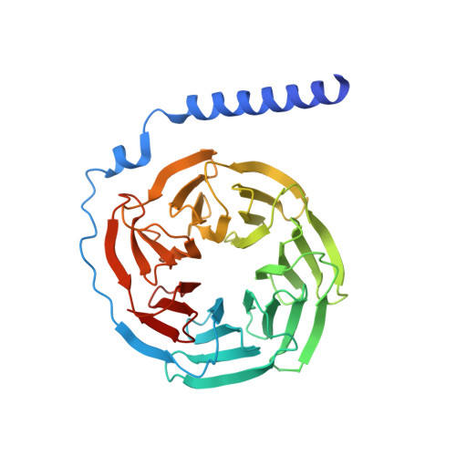
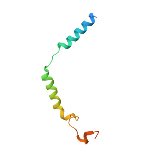
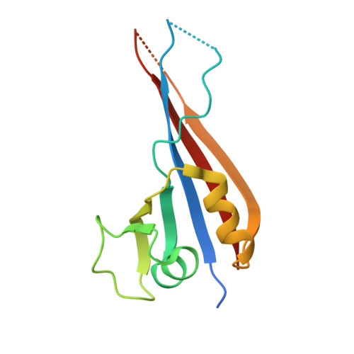
Structural basis for KCTD-mediated rapid desensitization of GABABsignalling.

Zheng, S.Abreu, N.Levitz, J.Kruse, A.C.

(2019) Nature 567: 127-131

  • DOI: https://doi.org/10.1038/s41586-019-0990-0
  • Primary Citation of Related Structures:  
    6M8R, 6M8S

  • PubMed Abstract: 

    The GABA B (γ-aminobutyric acid type B) receptor is one of the principal inhibitory neurotransmitter receptors in the brain, and it signals through heterotrimeric G proteins to activate a variety of effectors, including G-protein-coupled inwardly rectifying potassium channels (GIRKs) 1,2 . GABA B -receptor signalling is tightly regulated by auxiliary subunits called KCTDs, which control the kinetics of GIRK activation and desensitization 3-5 . However, the mechanistic basis for KCTD modulation of GABA B signalling remains incompletely understood. Here, using a combination of X-ray crystallography, electron microscopy, and functional and biochemical experiments, we reveal the molecular details of KCTD binding to both GABA B receptors and G-protein βγ subunits. KCTDs associate with the receptor by forming an asymmetric pentameric ring around a region of the receptor carboxy-terminal tail, while a second KCTD domain, H1, engages in a symmetric interaction with five copies of Gβγ in which the G-protein subunits also interact directly with one another. We further show that KCTD binding to Gβγ is highly cooperative, defining a model in which KCTD proteins cooperatively strip G proteins from GIRK channels to induce rapid desensitization following receptor activation. These results provide a framework for understanding the molecular basis for the precise temporal control of GABA B signalling by KCTD proteins.


  • Organizational Affiliation

    Department of Biological Chemistry and Molecular Pharmacology, Blavatnik Institute, Harvard Medical School, Boston, MA, USA.


Macromolecules
Find similar proteins by:  (by identity cutoff)  |  3D Structure
Entity ID: 1
MoleculeChains Sequence LengthOrganismDetailsImage
Guanine nucleotide-binding protein G(I)/G(S)/G(T) subunit beta-1A [auth C],
B [auth D],
C [auth G],
D [auth H],
G [auth K]
350Homo sapiensMutation(s): 0 
Gene Names: GNB1
UniProt & NIH Common Fund Data Resources
Find proteins for P62873 (Homo sapiens)
Explore P62873 
Go to UniProtKB:  P62873
PHAROS:  P62873
GTEx:  ENSG00000078369 
Entity Groups  
Sequence Clusters30% Identity50% Identity70% Identity90% Identity95% Identity100% Identity
UniProt GroupP62873
Sequence Annotations
Expand
  • Reference Sequence
Find similar proteins by:  (by identity cutoff)  |  3D Structure
Entity ID: 2
MoleculeChains Sequence LengthOrganismDetailsImage
Guanine nucleotide-binding protein G(I)/G(S)/G(O) subunit gamma-2E [auth I],
F [auth J],
H [auth L],
M [auth E],
N [auth F]
71Homo sapiensMutation(s): 0 
Gene Names: GNG2
UniProt & NIH Common Fund Data Resources
Find proteins for P59768 (Homo sapiens)
Explore P59768 
Go to UniProtKB:  P59768
PHAROS:  P59768
GTEx:  ENSG00000186469 
Entity Groups  
Sequence Clusters30% Identity50% Identity70% Identity90% Identity95% Identity100% Identity
UniProt GroupP59768
Sequence Annotations
Expand
  • Reference Sequence
Find similar proteins by:  (by identity cutoff)  |  3D Structure
Entity ID: 3
MoleculeChains Sequence LengthOrganismDetailsImage
BTB/POZ domain-containing protein KCTD12I [auth A],
J [auth O],
K [auth P],
L [auth B],
O [auth M]
129Homo sapiensMutation(s): 0 
Gene Names: KCTD12C13orf2KIAA1778PFET1
UniProt & NIH Common Fund Data Resources
Find proteins for Q96CX2 (Homo sapiens)
Explore Q96CX2 
Go to UniProtKB:  Q96CX2
PHAROS:  Q96CX2
GTEx:  ENSG00000178695 
Entity Groups  
Sequence Clusters30% Identity50% Identity70% Identity90% Identity95% Identity100% Identity
UniProt GroupQ96CX2
Sequence Annotations
Expand
  • Reference Sequence
Experimental Data & Validation

Experimental Data

  • Method: X-RAY DIFFRACTION
  • Resolution: 3.71 Å
  • R-Value Free: 0.287 
  • R-Value Work: 0.251 
  • R-Value Observed: 0.253 
  • Space Group: P 21 21 21
Unit Cell:
Length ( Å )Angle ( ˚ )
a = 109.09α = 90
b = 121.99β = 90
c = 206.43γ = 90
Software Package:
Software NamePurpose
PHENIXrefinement
XDSdata reduction
XDSdata scaling
PHASERphasing

Structure Validation

View Full Validation Report


Entry History & Funding Information

Deposition Data


Funding OrganizationLocationGrant Number
Other privateUnited StatesKlingenstein-Simons Fellowship Program

Revision History  (Full details and data files)

  • Version 1.0: 2019-02-27
    Type: Initial release
  • Version 1.1: 2019-03-13
    Changes: Data collection, Database references
  • Version 1.2: 2023-10-11
    Changes: Data collection, Database references, Refinement description