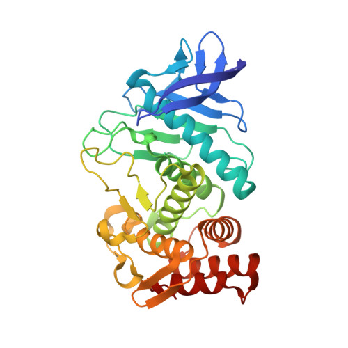
6LZN

Thermolysin

  • Classification: HYDROLASE
  • Organism(s): Bacillus thermoproteolyticus
  • Mutation(s): No 

  • Deposited: 2020-02-19 Released: 2021-01-27 
  • Deposition Author(s): Nam, K.H.
  • Funding Organization(s): National Research Foundation (NRF, Korea)

Experimental Data Snapshot

  • Method: X-RAY DIFFRACTION
  • Resolution: 1.50 Å
  • R-Value Free: 0.180 
  • R-Value Work: 0.157 
  • R-Value Observed: 0.158 

wwPDB Validation   3D Report Full Report


This is version 1.1 of the entry. See complete history


Literature

Structural analysis of metal chelation of the metalloproteinase thermolysin by 1,10-phenanthroline.

Nam, K.H.

(2021) J Inorg Biochem 215: 111319-111319

  • DOI: https://doi.org/10.1016/j.jinorgbio.2020.111319
  • Primary Citation of Related Structures:  
    6LZN, 6LZO

  • PubMed Abstract: 

    Metalloproteases and their inhibitors are important in numerous fundamental biochemical phenomena and medical applications. The heterocyclic organic compound, 1,10-phenanthroline, forms a complex with transition metal ions and is a Zn 2+ -chelating metalloprotease inhibitor; however, the mechanism of 1,10-phenanthroline-based chelation inhibition has not been fully elucidated. This study aimed to understand the structural basis of zinc metalloproteinase inhibition by 1,10-phenanthroline. Herein, the crystal structure of thermolysin was determined in the absence and presence of 1,10-phenanthroline at 1.5 and 1.8 Å, respectively. In native thermolysin, Zn 2+ at the active site is tetrahedrally coordinated by His142, His146, Glu166, and water molecule and contains three Ca 2+ ions, which are involved in thermostability. In the crystal structure of 1,10-phenanthroline-treated thermolysin crystal, seven 1,10-phenanthroline molecules were observed on the surface of thermolysin. These molecules are stabilized by π- π stacking interactions with aromatic amino acids (Phe63, Tyr66, Tyr110, His216, and Try251) or between the 1,10-phenanthrolines. Moreover, interactions with Ser5 and Arg101 were also observed. In this structure, Zn 2+ at the active site was completely chelated, but no large conformational changes were observed in Zn 2+ coordination with amino acid residues. Ca 2+ at the Ca3 site exposed to the solvent was chelated by 1,10-phenanthroline, resulting in a conformational change in the side chain of Asp56 and Gln61. Based on the surface structure, for 1,10-phenanthroline to chelate a metal, it is important that the metal is exposed on the protein surface and that there is no steric hindrance impairing 1,10-phenanthroline access by the amino acids around the metal.


  • Organizational Affiliation

    Department of Life Science, Pohang University of Science and Technology, Pohang 37673, Republic of Korea. Electronic address: structures@postech.ac.kr.


Macromolecules
Find similar proteins by:  (by identity cutoff)  |  3D Structure
Entity ID: 1
MoleculeChains Sequence LengthOrganismDetailsImage
Thermolysin316Bacillus thermoproteolyticusMutation(s): 0 
EC: 3.4.24.27
UniProt
Find proteins for P00800 (Bacillus thermoproteolyticus)
Explore P00800 
Go to UniProtKB:  P00800
Entity Groups  
Sequence Clusters30% Identity50% Identity70% Identity90% Identity95% Identity100% Identity
UniProt GroupP00800
Sequence Annotations
Expand
  • Reference Sequence
Small Molecules
Ligands 6 Unique
IDChains Name / Formula / InChI Key2D Diagram3D Interactions
LYS
Query on LYS

Download Ideal Coordinates CCD File 
I [auth A]LYSINE
C6 H15 N2 O2
KDXKERNSBIXSRK-YFKPBYRVSA-O
ILE
Query on ILE

Download Ideal Coordinates CCD File 
H [auth A]ISOLEUCINE
C6 H13 N O2
AGPKZVBTJJNPAG-WHFBIAKZSA-N
GOL
Query on GOL

Download Ideal Coordinates CCD File 
G [auth A]GLYCEROL
C3 H8 O3
PEDCQBHIVMGVHV-UHFFFAOYSA-N
ZN (Subject of Investigation/LOI)
Query on ZN

Download Ideal Coordinates CCD File 
B [auth A]ZINC ION
Zn
PTFCDOFLOPIGGS-UHFFFAOYSA-N
POL
Query on POL

Download Ideal Coordinates CCD File 
F [auth A]N-PROPANOL
C3 H8 O
BDERNNFJNOPAEC-UHFFFAOYSA-N
CA
Query on CA

Download Ideal Coordinates CCD File 
C [auth A],
D [auth A],
E [auth A]
CALCIUM ION
Ca
BHPQYMZQTOCNFJ-UHFFFAOYSA-N
Experimental Data & Validation

Experimental Data

  • Method: X-RAY DIFFRACTION
  • Resolution: 1.50 Å
  • R-Value Free: 0.180 
  • R-Value Work: 0.157 
  • R-Value Observed: 0.158 
  • Space Group: P 61 2 2
Unit Cell:
Length ( Å )Angle ( ˚ )
a = 92.663α = 90
b = 92.663β = 90
c = 128.591γ = 120
Software Package:
Software NamePurpose
REFMACrefinement
HKL-2000data scaling
PDB_EXTRACTdata extraction
HKL-2000data reduction
MOLREPphasing

Structure Validation

View Full Validation Report


Entry History & Funding Information

Deposition Data

  • Released Date: 2021-01-27 
  • Deposition Author(s): Nam, K.H.

Funding OrganizationLocationGrant Number
National Research Foundation (NRF, Korea)Korea, Republic OfNRF-2017R1D1A1B03033087
National Research Foundation (NRF, Korea)Korea, Republic OfNRF-2017M3A9F6029736

Revision History  (Full details and data files)

  • Version 1.0: 2021-01-27
    Type: Initial release
  • Version 1.1: 2023-11-29
    Changes: Data collection, Database references, Refinement description