6LOJ

The complex structure of IpaH9.8-LRR and hGBP1


Experimental Data Snapshot

  • Method: X-RAY DIFFRACTION
  • Resolution: 3.72 Å
  • R-Value Free: 0.256 
  • R-Value Work: 0.227 
  • R-Value Observed: 0.229 

wwPDB Validation   3D Report Full Report


Ligand Structure Quality Assessment 


This is version 1.2 of the entry. See complete history


Literature

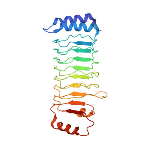
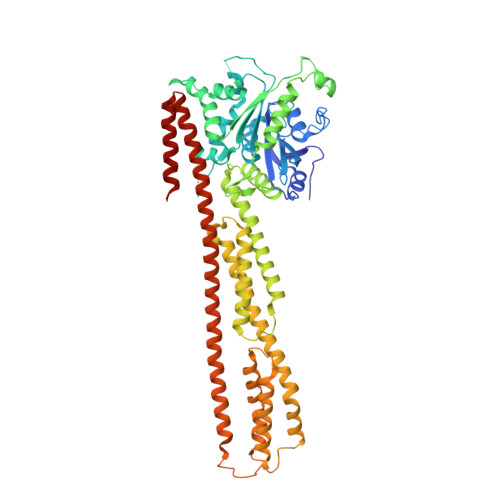
Substrate-binding destabilizes the hydrophobic cluster to relieve the autoinhibition of bacterial ubiquitin ligase IpaH9.8.

Ye, Y.Xiong, Y.Huang, H.

(2020) Commun Biol 3: 752-752

  • DOI: https://doi.org/10.1038/s42003-020-01492-1
  • Primary Citation of Related Structures:  
    6LOJ, 6LOL

  • PubMed Abstract: 

    IpaH enzymes are bacterial E3 ligases targeting host proteins for ubiquitylation. Two autoinhibition modes of IpaH enzymes have been proposed based on the relative positioning of the Leucine-rich repeat domain (LRR) with respect to the NEL domain. In mode 1, substrate-binding competitively displaces the interactions between theLRR and NEL to relieve autoinhibition. However, the molecular basis for mode 2 is unclear. Here, we present the crystal structures of Shigella IpaH9.8 and the LRR of IpaH9.8 in complex with the substrate of human guanylate-binding protein 1 (hGBP1). A hydrophobic cluster in the C-terminus of IpaH9.8 LRR forms a hydrophobic pocket involved in binding the NEL domain, and the binding is important for IpaH9.8 autoinhibition. Substrate-binding destabilizes the hydrophobic cluster by inducing conformational changes of IpaH9.8 LRR . Arg166 and Phe187 in IpaH9.8 LRR function as sensors for substrate-binding. Collectively, our findings provide insights into the molecular mechanisms for the actication of IpaH9.8 in autoinhibition mode 2.


  • Organizational Affiliation

    State Key Laboratory of Chemical Oncogenomics, School of Chemical Biology and Biotechnology, Peking University Shenzhen Graduate School, 518055, Shenzhen, China. yeyx@pkusz.edu.cn.


Macromolecules
Find similar proteins by:  (by identity cutoff)  |  3D Structure
Entity ID: 1
MoleculeChains Sequence LengthOrganismDetailsImage
Invasion plasmid antigen226Shigella flexneriMutation(s): 0 
Gene Names: ipaH9.8NCTC9783_05321
EC: 6.3.2
UniProt
Find proteins for D2AJU0 (Shigella flexneri serotype X (strain 2002017))
Explore D2AJU0 
Go to UniProtKB:  D2AJU0
Entity Groups  
Sequence Clusters30% Identity50% Identity70% Identity90% Identity95% Identity100% Identity
UniProt GroupD2AJU0
Sequence Annotations
Expand
  • Reference Sequence
Find similar proteins by:  (by identity cutoff)  |  3D Structure
Entity ID: 2
MoleculeChains Sequence LengthOrganismDetailsImage
Guanylate-binding protein 1595Homo sapiensMutation(s): 1 
Gene Names: GBP1
EC: 3.6.5
UniProt & NIH Common Fund Data Resources
Find proteins for P32455 (Homo sapiens)
Explore P32455 
Go to UniProtKB:  P32455
PHAROS:  P32455
GTEx:  ENSG00000117228 
Entity Groups  
Sequence Clusters30% Identity50% Identity70% Identity90% Identity95% Identity100% Identity
UniProt GroupP32455
Sequence Annotations
Expand
  • Reference Sequence
Small Molecules
Ligands 1 Unique
IDChains Name / Formula / InChI Key2D Diagram3D Interactions
GDP
Query on GDP

Download Ideal Coordinates CCD File 
C [auth B]GUANOSINE-5'-DIPHOSPHATE
C10 H15 N5 O11 P2
QGWNDRXFNXRZMB-UUOKFMHZSA-N
Experimental Data & Validation

Experimental Data

  • Method: X-RAY DIFFRACTION
  • Resolution: 3.72 Å
  • R-Value Free: 0.256 
  • R-Value Work: 0.227 
  • R-Value Observed: 0.229 
  • Space Group: P 61 2 2
Unit Cell:
Length ( Å )Angle ( ˚ )
a = 122.49α = 90
b = 122.49β = 90
c = 582.35γ = 120
Software Package:
Software NamePurpose
PHENIXrefinement
XSCALEdata scaling
PDB_EXTRACTdata extraction
XDSdata reduction
PHENIXphasing

Structure Validation

View Full Validation Report


Ligand Structure Quality Assessment 


Entry History & Funding Information

Deposition Data


Funding OrganizationLocationGrant Number
National Natural Science Foundation of China (NSFC)China21907006

Revision History  (Full details and data files)

  • Version 1.0: 2020-12-23
    Type: Initial release
  • Version 1.1: 2020-12-30
    Changes: Database references
  • Version 1.2: 2023-11-29
    Changes: Data collection, Database references, Refinement description