6L1H

Crystal structure of light-dependent protochlorophyllide oxidoreductase from Thermosynechococcus elongatus


Experimental Data Snapshot

  • Method: X-RAY DIFFRACTION
  • Resolution: 2.41 Å
  • R-Value Free: 0.221 
  • R-Value Work: 0.181 
  • R-Value Observed: 0.183 

wwPDB Validation   3D Report Full Report


Ligand Structure Quality Assessment 


This is version 1.3 of the entry. See complete history


Literature

Crystal structures of cyanobacterial light-dependent protochlorophyllide oxidoreductase.

Dong, C.S.Zhang, W.L.Wang, Q.Li, Y.S.Wang, X.Zhang, M.Liu, L.

(2020) Proc Natl Acad Sci U S A 117: 8455-8461

  • DOI: https://doi.org/10.1073/pnas.1920244117
  • Primary Citation of Related Structures:  
    6L1G, 6L1H

  • PubMed Abstract: 

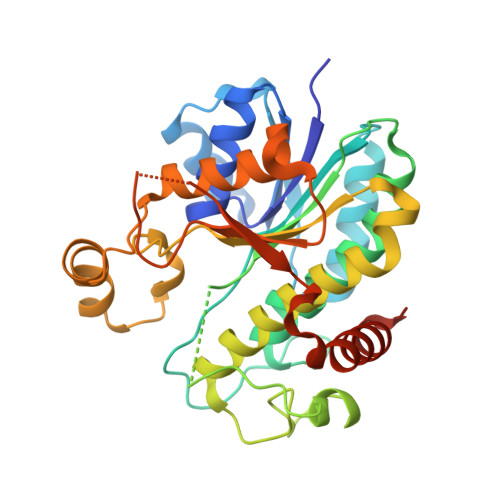
    The reduction of protochlorophyllide (Pchlide) to chlorophyllide (Chlide) is the penultimate step of chlorophyll biosynthesis. In oxygenic photosynthetic bacteria, algae, and plants, this reaction can be catalyzed by the light-dependent Pchlide oxidoreductase (LPOR), a member of the short-chain dehydrogenase superfamily sharing a conserved Rossmann fold for NAD(P)H binding and the catalytic activity. Whereas modeling and simulation approaches have been used to study the catalytic mechanism of this light-driven reaction, key details of the LPOR structure remain unclear. We determined the crystal structures of LPOR from two cyanobacteria, Synechocystis sp. PCC 6803 and Thermosynechococcus elongatus Structural analysis defines the LPOR core fold, outlines the LPOR-NADPH interaction network, identifies the residues forming the substrate cavity and the proton-relay path, and reveals the role of the LPOR-specific loop. These findings provide a basis for understanding the structure-function relationships of the light-driven Pchlide reduction.


  • Organizational Affiliation

    School of Life Sciences, Anhui University, 230601 Hefei, Anhui, China.


Macromolecules
Find similar proteins by:  (by identity cutoff)  |  3D Structure
Entity ID: 1
MoleculeChains Sequence LengthOrganismDetailsImage
NADPH-protochlorophyllide oxidoreductase
A, B
330Thermosynechococcus vestitus BP-1Mutation(s): 0 
Gene Names: por
EC: 1.3.1.33
UniProt
Find proteins for Q8DLC1 (Thermosynechococcus vestitus (strain NIES-2133 / IAM M-273 / BP-1))
Explore Q8DLC1 
Go to UniProtKB:  Q8DLC1
Entity Groups  
Sequence Clusters30% Identity50% Identity70% Identity90% Identity95% Identity100% Identity
UniProt GroupQ8DLC1
Sequence Annotations
Expand
  • Reference Sequence
Small Molecules
Ligands 1 Unique
IDChains Name / Formula / InChI Key2D Diagram3D Interactions
NDP (Subject of Investigation/LOI)
Query on NDP

Download Ideal Coordinates CCD File 
C [auth A],
D [auth B]
NADPH DIHYDRO-NICOTINAMIDE-ADENINE-DINUCLEOTIDE PHOSPHATE
C21 H30 N7 O17 P3
ACFIXJIJDZMPPO-NNYOXOHSSA-N
Experimental Data & Validation

Experimental Data

  • Method: X-RAY DIFFRACTION
  • Resolution: 2.41 Å
  • R-Value Free: 0.221 
  • R-Value Work: 0.181 
  • R-Value Observed: 0.183 
  • Space Group: P 21 21 21
Unit Cell:
Length ( Å )Angle ( ˚ )
a = 52.899α = 90
b = 96.2β = 90
c = 141.747γ = 90
Software Package:
Software NamePurpose
PHENIXrefinement
HKL-2000data reduction
HKL-2000data scaling
PHASERphasing
PDB_EXTRACTdata extraction

Structure Validation

View Full Validation Report


Ligand Structure Quality Assessment 


Entry History 

Deposition Data

Revision History  (Full details and data files)

  • Version 1.0: 2020-04-01
    Type: Initial release
  • Version 1.1: 2020-04-15
    Changes: Database references
  • Version 1.2: 2020-04-29
    Changes: Database references
  • Version 1.3: 2023-11-22
    Changes: Data collection, Database references, Refinement description