


Crystal structure of a standalone versatile EAL protein from Vibrio cholerae O395 - c-di-IMP bound form
Yadav, M., Pal, K., Sen, U.To be published.
Experimental Data Snapshot
Starting Model: experimental
View more details
Entity ID: 1 | |||||
|---|---|---|---|---|---|
| Molecule | Chains | Sequence Length | Organism | Details | Image |
| EAL domain protein | 257 | Vibrio cholerae O395 | Mutation(s): 1 Gene Names: VC0395_A1247 EC: 3.1.4.1 | 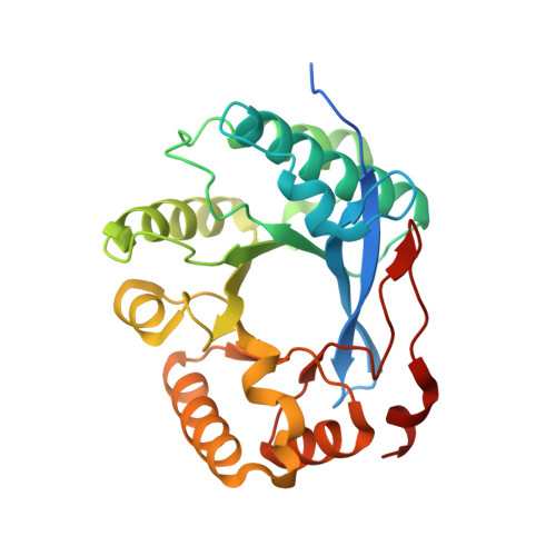 | |
UniProt | |||||
Find proteins for A0A0H3AJ04 (Vibrio cholerae serotype O1 (strain ATCC 39541 / Classical Ogawa 395 / O395)) Explore A0A0H3AJ04 Go to UniProtKB: A0A0H3AJ04 | |||||
Entity Groups | |||||
| Sequence Clusters | 30% Identity50% Identity70% Identity90% Identity95% Identity100% Identity | ||||
| UniProt Group | A0A0H3AJ04 | ||||
Sequence AnnotationsExpand | |||||
Reference Sequence | |||||
| Ligands 2 Unique | |||||
|---|---|---|---|---|---|
| ID | Chains | Name / Formula / InChI Key | 2D Diagram | 3D Interactions | |
| C2I (Subject of Investigation/LOI) Download:Ideal Coordinates CCD File | E [auth A], H [auth B], K [auth C], N [auth D] | 9-[(1R,6R,8R,9S,10R,15S,17R,18S)-3,9,12,18-tetrakis(oxidanyl)-3,12-bis(oxidanylidene)-17-(6-oxidanylidene-3H-purin-9-yl)-2,4,7,11,13,16-hexaoxa-3$l^{5},12$l^{5}-diphosphatricyclo[13.3.0.0^{6,10}]octadecan-8-yl]-3H-purin-6-one C20 H22 N8 O14 P2 VFTRASQVWRBMKD-PHSICLOESA-N |  | ||
| CA (Subject of Investigation/LOI) Download:Ideal Coordinates CCD File | F [auth A] G [auth A] I [auth B] J [auth B] L [auth C] | CALCIUM ION Ca BHPQYMZQTOCNFJ-UHFFFAOYSA-N |  | ||
| Length ( Å ) | Angle ( ˚ ) |
|---|---|
| a = 73.45 | α = 90 |
| b = 42.54 | β = 94.65 |
| c = 155.04 | γ = 90 |
| Software Name | Purpose |
|---|---|
| Aimless | data scaling |
| PHENIX | refinement |
| PDB_EXTRACT | data extraction |
| iMOSFLM | data reduction |
| PHENIX | phasing |


