6K2Q

Aeromonas hydrophila MtaN-2 complexed with adenine


Experimental Data Snapshot

  • Method: X-RAY DIFFRACTION
  • Resolution: 2.00 Å
  • R-Value Free: 0.195 
  • R-Value Work: 0.161 
  • R-Value Observed: 0.161 

wwPDB Validation   3D Report Full Report


Ligand Structure Quality Assessment 


This is version 1.2 of the entry. See complete history


Literature

Crystal Structure of Aeromonas hydrophila Cytoplasmic 5'-Methylthioadenosine/S-Adenosylhomocysteine Nucleosidase.

Chen, J.Liu, W.Wang, L.Shang, F.Chen, Y.Lan, J.Gao, P.Ha, N.C.Quan, C.Nam, K.H.Xu, Y.

(2019) Biochemistry 58: 3136-3143

  • DOI: https://doi.org/10.1021/acs.biochem.9b00174
  • Primary Citation of Related Structures:  
    6K2Q

  • PubMed Abstract: 

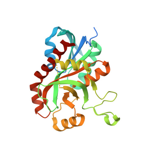
    5'-Methylthioadenosine/ S -adenosyl-l-homocysteine (MTA/SAH) nucleosidase (MTAN) is an important enzyme in a number of critical biological processes. Mammals do not express MtaN, making this enzyme an attractive antibacterial drug target. In pathogen Aeromonas hydrophila , two MtnN subfamily genes (MtaN-1 and MtaN-2) play important roles in the periplasm and cytosol, respectively. We previously reported structural and functional analyses of MtaN-1, but little is known regarding MtaN-2 due to the lack of a crystal structure. Here, we determined the crystal structure of cytosolic A. hydrophila MtaN-2 in complex with adenine (ADE), which is a cleavage product of adenosine. Ah MtaN-1 and Ah MtaN-2 exhibit a high degree of similarity in the α-β-α sandwich fold of the core structural motif. However, there is a structural difference in the nonconserved extended loop between β7 and α3 that is associated with the channel depth of the substrate-binding pocket and dimerization. The ADE molecules in the substrate-binding pockets of Ah MtaN-1 and Ah MtaN-2 are stabilized with π-π stacking by Trp199 and Phe152, respectively, and the hydrophobic residues surrounding the ribose-binding sites differ. A structural comparison of Ah MtaN-2 with other MtaN proteins showed that MtnN subfamily proteins exhibit a unique substrate-binding surface and dimerization interface.


  • Organizational Affiliation

    Department of Bioengineering, College of Life Science , Dalian Minzu University , Dalian 116600 , Liaoning , China.


Macromolecules
Find similar proteins by:  (by identity cutoff)  |  3D Structure
Entity ID: 1
MoleculeChains Sequence LengthOrganismDetailsImage
5'-methylthioadenosine/S-adenosylhomocysteine nucleosidase
A, B, C, D
260Aeromonas hydrophila subsp. hydrophila ATCC 7966Mutation(s): 0 
Gene Names: mtnNmtnN-2AHA_1706
EC: 3.2.2.9
UniProt
Find proteins for A0KIZ1 (Aeromonas hydrophila subsp. hydrophila (strain ATCC 7966 / DSM 30187 / BCRC 13018 / CCUG 14551 / JCM 1027 / KCTC 2358 / NCIMB 9240 / NCTC 8049))
Explore A0KIZ1 
Go to UniProtKB:  A0KIZ1
Entity Groups  
Sequence Clusters30% Identity50% Identity70% Identity90% Identity95% Identity100% Identity
UniProt GroupA0KIZ1
Sequence Annotations
Expand
  • Reference Sequence
Experimental Data & Validation

Experimental Data

  • Method: X-RAY DIFFRACTION
  • Resolution: 2.00 Å
  • R-Value Free: 0.195 
  • R-Value Work: 0.161 
  • R-Value Observed: 0.161 
  • Space Group: P 32
Unit Cell:
Length ( Å )Angle ( ˚ )
a = 74.939α = 90
b = 74.939β = 90
c = 185.214γ = 120
Software Package:
Software NamePurpose
PHENIXrefinement
HKL-2000data reduction
HKL-2000data scaling
PHENIXrefinement
PHENIXphasing

Structure Validation

View Full Validation Report


Ligand Structure Quality Assessment 


Entry History & Funding Information

Deposition Data


Funding OrganizationLocationGrant Number
National Natural Science Foundation of ChinaChina31200556
National Natural Science Foundation of ChinaChina21272031

Revision History  (Full details and data files)

  • Version 1.0: 2019-05-29
    Type: Initial release
  • Version 1.1: 2020-06-10
    Changes: Database references
  • Version 1.2: 2023-11-22
    Changes: Data collection, Database references, Refinement description