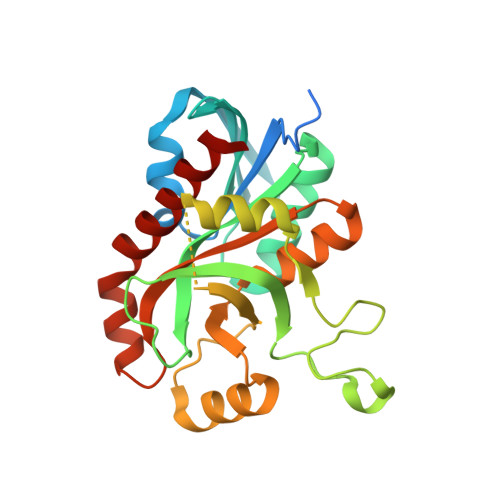
6K2Q

Aeromonas hydrophila MtaN-2 complexed with adenine


Experimental Data Snapshot

  • Method: X-RAY DIFFRACTION
  • Resolution: 2.00 Å
  • R-Value Free: 0.195 
  • R-Value Work: 0.161 
  • R-Value Observed: 0.161 

Starting Model: experimental
View more details

wwPDB Validation   3D Report Full Report


Ligand Structure Quality Assessment 


This is version 1.2 of the entry. See complete history


Literature

Crystal Structure of Aeromonas hydrophila Cytoplasmic 5'-Methylthioadenosine/S-Adenosylhomocysteine Nucleosidase.

Chen, J.Liu, W.Wang, L.Shang, F.Chen, Y.Lan, J.Gao, P.Ha, N.C.Quan, C.Nam, K.H.Xu, Y.

(2019) Biochemistry 58: 3136-3143

  • DOI: https://doi.org/10.1021/acs.biochem.9b00174
  • Primary Citation of Related Structures:  
    6K2Q

  • PubMed Abstract: 

    5'-Methylthioadenosine/ S -adenosyl-l-homocysteine (MTA/SAH) nucleosidase (MTAN) is an important enzyme in a number of critical biological processes. Mammals do not express MtaN, making this enzyme an attractive antibacterial drug target. In pathogen Aeromonas hydrophila , two MtnN subfamily genes (MtaN-1 and MtaN-2) play important roles in the periplasm and cytosol, respectively. We previously reported structural and functional analyses of MtaN-1, but little is known regarding MtaN-2 due to the lack of a crystal structure. Here, we determined the crystal structure of cytosolic A. hydrophila MtaN-2 in complex with adenine (ADE), which is a cleavage product of adenosine. Ah MtaN-1 and Ah MtaN-2 exhibit a high degree of similarity in the α-β-α sandwich fold of the core structural motif. However, there is a structural difference in the nonconserved extended loop between β7 and α3 that is associated with the channel depth of the substrate-binding pocket and dimerization. The ADE molecules in the substrate-binding pockets of Ah MtaN-1 and Ah MtaN-2 are stabilized with π-π stacking by Trp199 and Phe152, respectively, and the hydrophobic residues surrounding the ribose-binding sites differ. A structural comparison of Ah MtaN-2 with other MtaN proteins showed that MtnN subfamily proteins exhibit a unique substrate-binding surface and dimerization interface.


  • Organizational Affiliation

    Department of Bioengineering, College of Life Science , Dalian Minzu University , Dalian 116600 , Liaoning , China.


Macromolecules
Find similar proteins by:  (by identity cutoff)  |  3D Structure
Entity ID: 1
MoleculeChains Sequence LengthOrganismDetailsImage
5'-methylthioadenosine/S-adenosylhomocysteine nucleosidase
A, B, C, D
260Aeromonas hydrophila subsp. hydrophila ATCC 7966Mutation(s): 0 
Gene Names: mtnNmtnN-2AHA_1706
EC: 3.2.2.9
UniProt
Find proteins for A0KIZ1 (Aeromonas hydrophila subsp. hydrophila (strain ATCC 7966 / DSM 30187 / BCRC 13018 / CCUG 14551 / JCM 1027 / KCTC 2358 / NCIMB 9240 / NCTC 8049))
Explore A0KIZ1 
Go to UniProtKB:  A0KIZ1
Entity Groups  
Sequence Clusters30% Identity50% Identity70% Identity90% Identity95% Identity100% Identity
UniProt GroupA0KIZ1
Sequence Annotations
Expand
  • Reference Sequence
Experimental Data & Validation

Experimental Data

  • Method: X-RAY DIFFRACTION
  • Resolution: 2.00 Å
  • R-Value Free: 0.195 
  • R-Value Work: 0.161 
  • R-Value Observed: 0.161 
  • Space Group: P 32
Unit Cell:
Length ( Å )Angle ( ˚ )
a = 74.939α = 90
b = 74.939β = 90
c = 185.214γ = 120
Software Package:
Software NamePurpose
PHENIXrefinement
HKL-2000data reduction
HKL-2000data scaling
PHENIXrefinement
PHENIXphasing

Structure Validation

View Full Validation Report


Ligand Structure Quality Assessment 


Entry History & Funding Information

Deposition Data


Funding OrganizationLocationGrant Number
National Natural Science Foundation of ChinaChina31200556
National Natural Science Foundation of ChinaChina21272031

Revision History  (Full details and data files)

  • Version 1.0: 2019-05-29
    Type: Initial release
  • Version 1.1: 2020-06-10
    Changes: Database references
  • Version 1.2: 2023-11-22
    Changes: Data collection, Database references, Refinement description