6K2D

The crystal structure of GBP1 with LRR domain of IpaH9.8


Experimental Data Snapshot

  • Method: X-RAY DIFFRACTION
  • Resolution: 3.60 Å
  • R-Value Free: 0.287 
  • R-Value Work: 0.240 
  • R-Value Observed: 0.243 

wwPDB Validation   3D Report Full Report


This is version 1.2 of the entry. See complete history


Literature

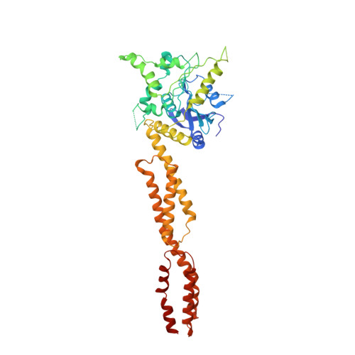
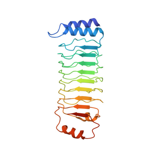
Structural mechanism for guanylate-binding proteins (GBPs) targeting by the Shigella E3 ligase IpaH9.8.

Ji, C.Du, S.Li, P.Zhu, Q.Yang, X.Long, C.Yu, J.Shao, F.Xiao, J.Y.

(2019) PLoS Pathog 15: e1007876-e1007876

  • DOI: https://doi.org/10.1371/journal.ppat.1007876
  • Primary Citation of Related Structures:  
    6K1Z, 6K2D

  • PubMed Abstract: 

    The guanylate-binding proteins (GBPs) belong to the dynamin superfamily of GTPases and function in cell-autonomous defense against intracellular pathogens. IpaH9.8, an E3 ligase from the pathogenic bacterium Shigella flexneri, ubiquitinates a subset of GBPs and leads to their proteasomal degradation. Here we report the structure of a C-terminally truncated GBP1 in complex with the IpaH9.8 Leucine-rich repeat (LRR) domain. IpaH9.8LRR engages the GTPase domain of GBP1, and differences in the Switch II and α3 helix regions render some GBPs such as GBP3 and GBP7 resistant to IpaH9.8. Comparisons with other IpaH structures uncover interaction hot spots in their LRR domains. The C-terminal region of GBP1 undergoes a large rotation compared to previously determined structures. We further show that the C-terminal farnesylation modification also plays a role in regulating GBP1 conformation. Our results suggest a general mechanism by which the IpaH proteins target their cellular substrates and shed light on the structural dynamics of the GBPs.


  • Organizational Affiliation

    The State Key Laboratory of Protein and Plant Gene Research, School of Life Sciences, Peking-Tsinghua Center for Life Sciences, Peking University, Beijing, China.


Macromolecules
Find similar proteins by:  (by identity cutoff)  |  3D Structure
Entity ID: 1
MoleculeChains Sequence LengthOrganismDetailsImage
Guanylate-binding protein 1481Homo sapiensMutation(s): 0 
Gene Names: GBP1
EC: 3.6.5
UniProt & NIH Common Fund Data Resources
Find proteins for P32455 (Homo sapiens)
Explore P32455 
Go to UniProtKB:  P32455
PHAROS:  P32455
GTEx:  ENSG00000117228 
Entity Groups  
Sequence Clusters30% Identity50% Identity70% Identity90% Identity95% Identity100% Identity
UniProt GroupP32455
Sequence Annotations
Expand
  • Reference Sequence
Find similar proteins by:  (by identity cutoff)  |  3D Structure
Entity ID: 2
MoleculeChains Sequence LengthOrganismDetailsImage
E3 ubiquitin-protein ligase ipaH9.8236Shigella flexneriMutation(s): 0 
Gene Names: ipaH9.8CP0226pWR501_0234SFLP090
EC: 2.3.2.27
UniProt
Find proteins for Q8VSC3 (Shigella flexneri)
Explore Q8VSC3 
Go to UniProtKB:  Q8VSC3
Entity Groups  
Sequence Clusters30% Identity50% Identity70% Identity90% Identity95% Identity100% Identity
UniProt GroupQ8VSC3
Sequence Annotations
Expand
  • Reference Sequence
Experimental Data & Validation

Experimental Data

  • Method: X-RAY DIFFRACTION
  • Resolution: 3.60 Å
  • R-Value Free: 0.287 
  • R-Value Work: 0.240 
  • R-Value Observed: 0.243 
  • Space Group: P 31 2 1
Unit Cell:
Length ( Å )Angle ( ˚ )
a = 211.736α = 90
b = 211.736β = 90
c = 56.975γ = 120
Software Package:
Software NamePurpose
PHENIXrefinement
HKL-2000data collection
HKL-2000data scaling
PHENIXphasing

Structure Validation

View Full Validation Report


Entry History 

Deposition Data

Revision History  (Full details and data files)

  • Version 1.0: 2019-06-12
    Type: Initial release
  • Version 1.1: 2019-12-25
    Changes: Database references
  • Version 1.2: 2024-03-27
    Changes: Data collection, Database references