6JUE

The complex of PDZ and PBM


Experimental Data Snapshot

  • Method: X-RAY DIFFRACTION
  • Resolution: 1.55 Å
  • R-Value Free: 0.223 
  • R-Value Work: 0.211 
  • R-Value Observed: 0.211 

wwPDB Validation   3D Report Full Report


This is version 1.2 of the entry. See complete history


Literature

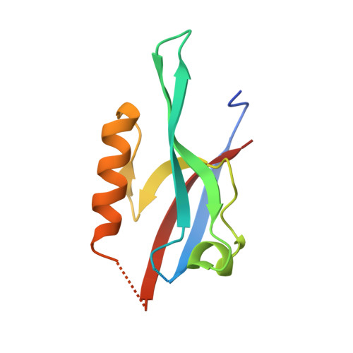
Par complex cluster formation mediated by phase separation.

Liu, Z.Yang, Y.Gu, A.Xu, J.Mao, Y.Lu, H.Hu, W.Lei, Q.Y.Li, Z.Zhang, M.Cai, Y.Wen, W.

(2020) Nat Commun 11: 2266-2266

  • DOI: https://doi.org/10.1038/s41467-020-16135-6
  • Primary Citation of Related Structures:  
    6JUE

  • PubMed Abstract: 

    The evolutionarily conserved Par3/Par6/aPKC complex regulates the polarity establishment of diverse cell types and distinct polarity-driven functions. However, how the Par complex is concentrated beneath the membrane to initiate cell polarization remains unclear. Here we show that the Par complex exhibits cell cycle-dependent condensation in Drosophila neuroblasts, driven by liquid-liquid phase separation. The open conformation of Par3 undergoes autonomous phase separation likely due to its NTD-mediated oligomerization. Par6, via C-terminal tail binding to Par3 PDZ3, can be enriched to Par3 condensates and in return dramatically promote Par3 phase separation. aPKC can also be concentrated to the Par3N/Par6 condensates as a client. Interestingly, activated aPKC can disperse the Par3/Par6 condensates via phosphorylation of Par3. Perturbations of Par3/Par6 phase separation impair the establishment of apical-basal polarity during neuroblast asymmetric divisions and lead to defective lineage development. We propose that phase separation may be a common mechanism for localized cortical condensation of cell polarity complexes.


  • Organizational Affiliation

    Department of Neurosurgery, Huashan Hospital, the Shanghai Key Laboratory of Medical Epigenetics, the International Co-laboratory of Medical Epigenetics and Metabolism, Ministry of Science and Technology, Institutes of Biomedical Sciences, State Key Laboratory of Medical Neurobiology and MOE Frontiers Center for Brain Science, School of Basic Medical Sciences, Fudan University, Shanghai, 200032, China.


Macromolecules
Find similar proteins by:  (by identity cutoff)  |  3D Structure
Entity ID: 1
MoleculeChains Sequence LengthOrganismDetailsImage
Partitioning defective 3 homologA [auth L]110Rattus norvegicusMutation(s): 0 
UniProt
Find proteins for Q9Z340 (Rattus norvegicus)
Explore Q9Z340 
Go to UniProtKB:  Q9Z340
Entity Groups  
Sequence Clusters30% Identity50% Identity70% Identity90% Identity95% Identity100% Identity
UniProt GroupQ9Z340
Sequence Annotations
Expand
  • Reference Sequence

Find similar proteins by:  Sequence   |   3D Structure  

Entity ID: 2
MoleculeChains Sequence LengthOrganismDetailsImage
THR-ILE-ILE-THR-LEUB [auth A]10Mus musculusMutation(s): 0 
UniProt
Find proteins for Q9JK83 (Mus musculus)
Explore Q9JK83 
Go to UniProtKB:  Q9JK83
Entity Groups  
Sequence Clusters30% Identity50% Identity70% Identity90% Identity95% Identity100% Identity
UniProt GroupQ9JK83
Sequence Annotations
Expand
  • Reference Sequence
Experimental Data & Validation

Experimental Data

  • Method: X-RAY DIFFRACTION
  • Resolution: 1.55 Å
  • R-Value Free: 0.223 
  • R-Value Work: 0.211 
  • R-Value Observed: 0.211 
  • Space Group: P 63
Unit Cell:
Length ( Å )Angle ( ˚ )
a = 57.454α = 90
b = 57.454β = 90
c = 52.313γ = 120
Software Package:
Software NamePurpose
PHENIXrefinement
XDSdata reduction
autoPROCdata scaling
PHENIXphasing

Structure Validation

View Full Validation Report


Entry History & Funding Information

Deposition Data

  • Released Date: 2020-04-22 
  • Deposition Author(s): Liu, Z.

Funding OrganizationLocationGrant Number
National Natural Science Foundation of ChinaChina31871394
National Natural Science Foundation of ChinaChina31670730
National Natural Science Foundation of ChinaChina31422015
Ministry of Science and Technology (China)China2014CB910201

Revision History  (Full details and data files)

  • Version 1.0: 2020-04-22
    Type: Initial release
  • Version 1.1: 2020-11-04
    Changes: Database references, Refinement description, Structure summary
  • Version 1.2: 2023-11-22
    Changes: Data collection, Database references, Refinement description