Molecular and Structural Basis of the Proteasome alpha Subunit Assembly Mechanism Mediated by the Proteasome-Assembling Chaperone PAC3-PAC4 Heterodimer.
Satoh, T., Yagi-Utsumi, M., Okamoto, K., Kurimoto, E., Tanaka, K., Kato, K.(2019) Int J Mol Sci 20
- PubMed: 31067643 Search on PubMedSearch on PubMed Central
- DOI: https://doi.org/10.3390/ijms20092231
- Primary Citation Related Structures:
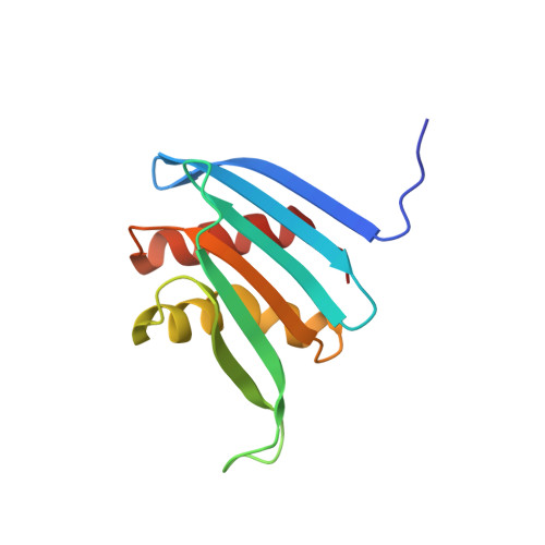
6JPT - PubMed Abstract:
The 26S proteasome is critical for the selective degradation of proteins in eukaryotic cells. This enzyme complex is composed of approximately 70 subunits, including the structurally homologous proteins α1-α7, which combine to form heptameric rings. The correct arrangement of these α subunits is essential for the function of the proteasome, but their assembly does not occur autonomously. Assembly of the α subunit is assisted by several chaperones, including the PAC3-PAC4 heterodimer. In this study we showed that the PAC3-PAC4 heterodimer functions as a molecular matchmaker, stabilizing the α4-α5-α6 subcomplex during the assembly of the α-ring. We solved a 0.96-Å atomic resolution crystal structure for a PAC3 homodimer which, in conjunction with nuclear magnetic resonance (NMR) data, highlighted the mobility of the loop comprised of residues 51 to 61. Based on these structural and dynamic data, we created a three-dimensional model of the PAC3-4/α4/α5/α6 quintet complex, and used this model to investigate the molecular and structural basis of the mechanism of proteasome α subunit assembly, as mediated by the PAC3-PAC4 heterodimeric chaperone. Our results provide a potential basis for the development of selective inhibitors against proteasome biogenesis.
- Graduate School of Pharmaceutical Sciences, Nagoya City University, 3-1 Tanabe-dori, Mizuho-ku, Nagoya 467-8603, Japan. tadashisatoh@phar.nagoya-cu.ac.jp.
Organizational Affiliation: