6JPR

Crystal structure of Phycocyanin from Nostoc sp. R76DM


Experimental Data Snapshot

  • Method: X-RAY DIFFRACTION
  • Resolution: 2.35 Å
  • R-Value Free: 0.248 
  • R-Value Work: 0.199 
  • R-Value Observed: 0.201 

wwPDB Validation   3D Report Full Report


Ligand Structure Quality Assessment 


This is version 1.3 of the entry. See complete history


Literature

Phylogenetic and crystallographic analysis of Nostoc phycocyanin having blue-shifted spectral properties.

Sonani, R.R.Rastogi, R.P.Patel, S.N.Chaubey, M.G.Singh, N.K.Gupta, G.D.Kumar, V.Madamwar, D.

(2019) Sci Rep 9: 9863-9863

  • DOI: https://doi.org/10.1038/s41598-019-46288-4
  • Primary Citation of Related Structures:  
    6JPR

  • PubMed Abstract: 

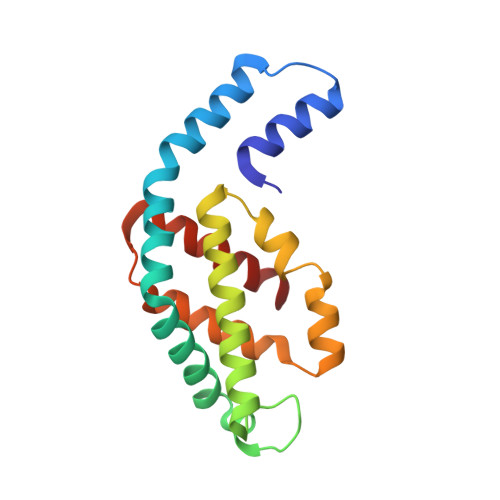
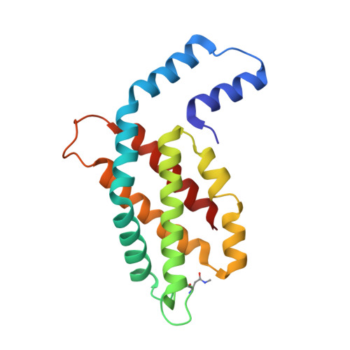
    The distinct sequence feature and spectral blue-shift (~10 nm) of phycocyanin, isolated from Nostoc sp. R76DM (N-PC), were investigated by phylogenetic and crystallographic analyses. Twelve conserved substitutions in N-PC sequence were found distributed unequally among α- and β-subunit (3 in α- and 9 in β-subunit). The phylogenetic analysis suggested that molecular evolution of α- and β-subunit of Nostoc-phycocyanin is faster than evolution of Nostoc-species. The divergence events seem to have occurred more frequently in β-subunit, compared to α-subunit (relative divergence, 7.38 for α-subunit and 9.66 for β-subunit). Crystal structure of N-PC was solved at 2.35 Å resolution to reasonable R-factors (R work /R Free  = 0.199/0.248). Substitutions congregate near interface of two αβ-monomer in N-PC trimer and are of compensatory nature. Six of the substitutions in β-subunit may be involved in maintaining topology of β-subunit, one in inter-monomer interaction and one in interaction with linker-protein. The β153Cys-attached chromophore adopts high-energy conformational state resulting due to reduced coplanarity of B- and C-pyrrole rings. Distortion in chromophore conformation can result in blue-shift in N-PC spectral properties. N-PC showed significant in-vitro and in-vivo antioxidant activity comparable with other phycocyanin. Since Nostoc-species constitute a distinct phylogenetic clade, the present structure would provide a better template to build a model for phycocyanins of these species.


  • Organizational Affiliation

    Radiation Biology & Health Sciences Division, Bhabha Atomic Research Centre, Trombay, Mumbai, 400 085, India.


Macromolecules
Find similar proteins by:  (by identity cutoff)  |  3D Structure
Entity ID: 1
MoleculeChains Sequence LengthOrganismDetailsImage
Phycocyanin
A, C, E, G, I
A, C, E, G, I, K
162NostocMutation(s): 0 
UniProt
Find proteins for A0A5H1ZR40 (Nostoc)
Explore A0A5H1ZR40 
Go to UniProtKB:  A0A5H1ZR40
Entity Groups  
Sequence Clusters30% Identity50% Identity70% Identity90% Identity95% Identity100% Identity
UniProt GroupA0A5H1ZR40
Sequence Annotations
Expand
  • Reference Sequence
Find similar proteins by:  (by identity cutoff)  |  3D Structure
Entity ID: 2
MoleculeChains Sequence LengthOrganismDetailsImage
Phycocyanin
B, D, F, H, J
B, D, F, H, J, L
172NostocMutation(s): 0 
UniProt
Find proteins for A0A5H1ZR41 (Nostoc)
Explore A0A5H1ZR41 
Go to UniProtKB:  A0A5H1ZR41
Entity Groups  
Sequence Clusters30% Identity50% Identity70% Identity90% Identity95% Identity100% Identity
UniProt GroupA0A5H1ZR41
Sequence Annotations
Expand
  • Reference Sequence
Small Molecules
Ligands 3 Unique
IDChains Name / Formula / InChI Key2D Diagram3D Interactions
CYC (Subject of Investigation/LOI)
Query on CYC

Download Ideal Coordinates CCD File 
AA [auth E]
CA [auth F]
DA [auth F]
FA [auth G]
HA [auth H]
AA [auth E],
CA [auth F],
DA [auth F],
FA [auth G],
HA [auth H],
IA [auth H],
LA [auth I],
M [auth A],
MA [auth J],
NA [auth J],
P [auth B],
Q [auth B],
QA [auth K],
SA [auth L],
T [auth C],
TA [auth L],
V [auth D],
W [auth D]
PHYCOCYANOBILIN
C33 H40 N4 O6
VXTXPYZGDQPMHK-GMXXPEQVSA-N
GOL
Query on GOL

Download Ideal Coordinates CCD File 
BA [auth E]
GA [auth G]
JA [auth H]
KA [auth H]
OA [auth J]
BA [auth E],
GA [auth G],
JA [auth H],
KA [auth H],
OA [auth J],
PA [auth J],
R [auth B],
RA [auth K],
U [auth C],
X [auth D],
Y [auth D]
GLYCEROL
C3 H8 O3
PEDCQBHIVMGVHV-UHFFFAOYSA-N
NA
Query on NA

Download Ideal Coordinates CCD File 
EA [auth F],
N [auth A],
O [auth A],
S [auth B],
Z [auth D]
SODIUM ION
Na
FKNQFGJONOIPTF-UHFFFAOYSA-N
Modified Residues  1 Unique
IDChains TypeFormula2D DiagramParent
MEN
Query on MEN
B, D, F, H, J
B, D, F, H, J, L
L-PEPTIDE LINKINGC5 H10 N2 O3ASN
Experimental Data & Validation

Experimental Data

  • Method: X-RAY DIFFRACTION
  • Resolution: 2.35 Å
  • R-Value Free: 0.248 
  • R-Value Work: 0.199 
  • R-Value Observed: 0.201 
  • Space Group: P 1 21 1
Unit Cell:
Length ( Å )Angle ( ˚ )
a = 67.136α = 90
b = 186.158β = 94.33
c = 85.532γ = 90
Software Package:
Software NamePurpose
PHENIXrefinement
XDSdata reduction
Aimlessdata scaling
PHASERphasing

Structure Validation

View Full Validation Report


Ligand Structure Quality Assessment 


Entry History & Funding Information

Deposition Data


Funding OrganizationLocationGrant Number
Department of Science & Technology (India)IndiaPDF.2017.001596

Revision History  (Full details and data files)

  • Version 1.0: 2019-06-26
    Type: Initial release
  • Version 1.1: 2019-07-17
    Changes: Data collection, Database references
  • Version 1.2: 2019-07-24
    Changes: Data collection, Database references
  • Version 1.3: 2023-11-22
    Changes: Data collection, Database references, Derived calculations, Refinement description