


Structure of a triple-helix region of human collagen type II.
Yang, X., Zhu, Y., Ye, S., Zhang, R., Lu, L.To be published.
Experimental Data Snapshot
wwPDB Validation 3D Report Full Report

Entity ID: 1 | |||||
|---|---|---|---|---|---|
| Molecule | Chains | Sequence Length | Organism | Details | Image |
| A triple-helix region of human collagen type II | 29 | Homo sapiens | Mutation(s): 0 | 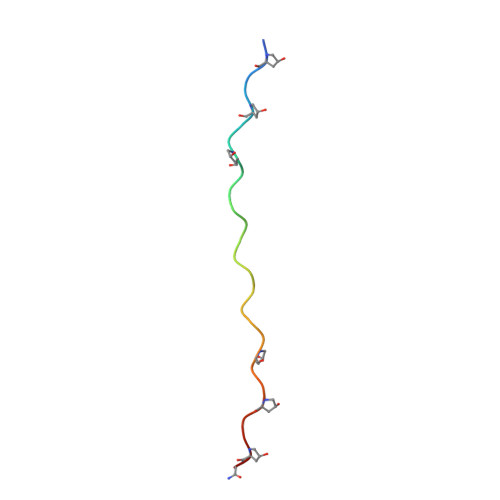 | |
Entity Groups | |||||
| Sequence Clusters | 30% Identity50% Identity70% Identity90% Identity95% Identity100% Identity | ||||
Sequence AnnotationsExpand | |||||
Reference Sequence | |||||
| Modified Residues 1 Unique | |||||
|---|---|---|---|---|---|
| ID | Chains | Type | Formula | 2D Diagram | Parent |
| HYP Query on HYP | A, B, C | L-PEPTIDE LINKING | C5 H9 N O3 |  | PRO |
| Length ( Å ) | Angle ( ˚ ) |
|---|---|
| a = 60.541 | α = 90 |
| b = 17.424 | β = 114.39 |
| c = 51.637 | γ = 90 |
| Software Name | Purpose |
|---|---|
| PHENIX | refinement |
| HKL-2000 | data reduction |
| HKL-2000 | data scaling |
| PHASER | phasing |


