6JJZ | pdb_00006jjz

Crystal Structure of MAGI2 and Dendrin complex


Experimental Data Snapshot

  • Method: X-RAY DIFFRACTION
  • Resolution: 1.65 Å
  • R-Value Free: 
    0.238 (Depositor), 0.239 (DCC) 
  • R-Value Work: 
    0.204 (Depositor), 0.206 (DCC) 
  • R-Value Observed: 
    0.206 (Depositor) 

Starting Model: experimental
View more details

wwPDB Validation 3D Report Full Report

Validation slider image for 6JJZ

This is version 1.1 of the entry. See complete history

Literature

Decoding WW domain tandem-mediated target recognitions in tissue growth and cell polarity.

Lin, Z.Yang, Z.Xie, R.Ji, Z.Guan, K.Zhang, M.

(2019) Elife 8

  • DOI: https://doi.org/10.7554/eLife.49439
  • Primary Citation Related Structures: 
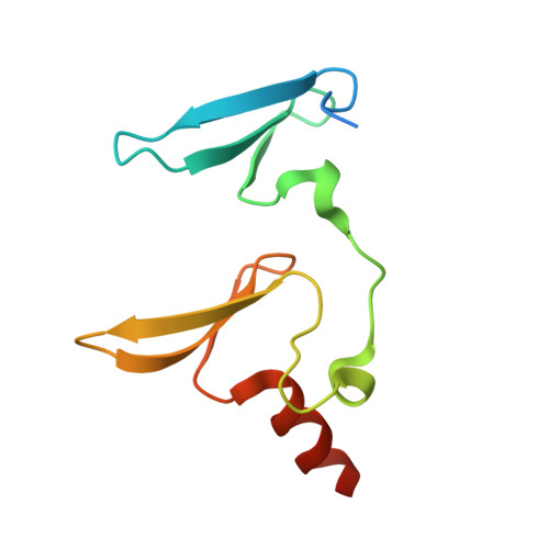
    6J68, 6JJW, 6JJX, 6JJY, 6JJZ, 6JK0, 6JK1

  • PubMed Abstract: 

    WW domain tandem-containing proteins such as KIBRA, YAP, and MAGI play critical roles in cell growth and polarity via binding to and positioning target proteins in specific subcellular regions. An immense disparity exists between promiscuity of WW domain-mediated target bindings and specific roles of WW domain proteins in cell growth regulation. Here, we discovered that WW domain tandems of KIBRA and MAGI, but not YAP, bind to specific target proteins with extremely high affinity and exquisite sequence specificity. Via systematic structural biology and biochemistry approaches, we decoded the target binding rules of WW domain tandems from cell growth regulatory proteins and uncovered a list of previously unknown WW tandem binding proteins including β-Dystroglycan, JCAD, and PTPN21. The WW tandem-mediated target recognition mechanisms elucidated here can guide functional studies of WW domain proteins in cell growth and polarity as well as in other cellular processes including neuronal synaptic signaling.


  • Organizational Affiliation
    • Division of Life Science, State Key Laboratory of Molecular Neuroscience, Hong Kong University of Science and Technology, Hong Kong, China.

Macromolecule Content 

  • Total Structure Weight: 28.08 kDa 
  • Atom Count: 1,850 
  • Modeled Residue Count: 210 
  • Deposited Residue Count: 240 
  • Unique protein chains: 2

Macromolecules

Find similar proteins by:|  3D Structure
Entity ID: 1
MoleculeChains  Sequence LengthOrganismDetailsImage
Membrane-associated guanylate kinase, WW and PDZ domain-containing protein 2
A, B
102Mus musculusMutation(s): 0 
Gene Names: Magi2Acvrinp1Aip1Arip1
UniProt & NIH Common Fund Data Resources
Find proteins for Q9WVQ1 (Mus musculus)
Explore Q9WVQ1 
Go to UniProtKB:  Q9WVQ1
IMPC:  MGI:1354953
Entity Groups
Sequence Clusters30% Identity50% Identity70% Identity90% Identity95% Identity100% Identity
UniProt GroupQ9WVQ1
Sequence Annotations
Expand
Reference Sequence
Find similar proteins by:  Sequence   |   3D Structure  
Entity ID: 2
MoleculeChains  Sequence LengthOrganismDetailsImage
Peptide from Dendrin
C, D
18Mus musculusMutation(s): 0 
Gene Names: DdnGm748Kiaa0749
UniProt
Find proteins for Q80TS7 (Mus musculus)
Explore Q80TS7 
Go to UniProtKB:  Q80TS7
Entity Groups
Sequence Clusters30% Identity50% Identity70% Identity90% Identity95% Identity100% Identity
UniProt GroupQ80TS7
Sequence Annotations
Expand
Reference Sequence

Experimental Data & Validation

Experimental Data

  • Method: X-RAY DIFFRACTION
  • Resolution: 1.65 Å
  • R-Value Free:  0.238 (Depositor), 0.239 (DCC) 
  • R-Value Work:  0.204 (Depositor), 0.206 (DCC) 
  • R-Value Observed: 0.206 (Depositor) 
Space Group: P 21 21 21
Unit Cell:
Length ( Å )Angle ( ˚ )
a = 46.899α = 90
b = 58.582β = 90
c = 86.298γ = 90
Software Package:
Software NamePurpose
PHENIXrefinement
HKL-2000data reduction
HKL-2000data scaling
PHASERphasing

Structure Validation

View Full Validation Report


Entry History 

Deposition Data

Revision History  (Full details and data files)

  • Version 1.0: 2019-09-25
    Type: Initial release
  • Version 1.1: 2023-11-22
    Changes: Data collection, Database references, Refinement description