Convergent evolution of the Cys decarboxylases involved in aminovinyl-cysteine (AviCys) biosynthesis.
Mo, T., Yuan, H., Wang, F., Ma, S., Wang, J., Li, T., Liu, G., Yu, S., Tan, X., Ding, W., Zhang, Q.(2019) FEBS Lett 593: 573-580
- PubMed: 30771247 Search on PubMed
- DOI: https://doi.org/10.1002/1873-3468.13341
- Primary Citation Related Structures:
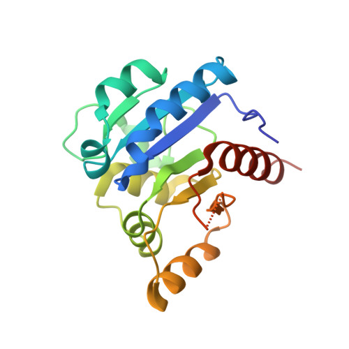
6JDD - PubMed Abstract:
S-[(Z)-2-aminovinyl]-d-cysteine (AviCys) is a unique motif found in several classes of ribosomally synthesized and post-translationally modified peptides (RiPPs). Biosynthesis of AviCys requires flavin-dependent Cys decarboxylases, which are highly divergent among different RiPP classes. In this study, we solved the crystal structure of the cypemycin decarboxylase CypD. We show that CypD is structurally highly similar to lanthipeptide decarboxylases despite the absence of sequence similarities between them. We further show that Cys decarboxylases from four RiPP classes have evolved independently and form two major clusters. These results reveal the convergent evolution of AviCys biosynthesis and suggest that all the flavin-dependent Cys decarboxylases likely have a similar Rossmann fold despite their sequence divergences.
- Department of Chemistry, Fudan University, Shanghai, China.
Organizational Affiliation: