6J95 | pdb_00006j95

Crystal structure of CYP97A3 in complex with retinal


Experimental Data Snapshot

  • Method: X-RAY DIFFRACTION
  • Resolution: 2.00 Å
  • R-Value Free: 
    0.197 (Depositor), 0.198 (DCC) 
  • R-Value Work: 
    0.165 (Depositor), 0.168 (DCC) 
  • R-Value Observed: 
    0.167 (Depositor) 

Starting Model: experimental
View more details

wwPDB Validation 3D Report Full Report

Validation slider image for 6J95

Ligand Structure Quality Assessment 


This is version 1.4 of the entry. See complete history

Literature

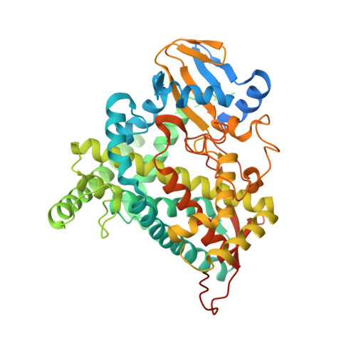
Structural basis for plant lutein biosynthesis from alpha-carotene.

Niu, G.Guo, Q.Wang, J.Zhao, S.He, Y.Liu, L.

(2020) Proc Natl Acad Sci U S A 117: 14150-14157

  • DOI: https://doi.org/10.1073/pnas.2001806117
  • Primary Citation Related Structures: 
    6J94, 6J95, 6L8H, 6L8I, 6L8J

  • PubMed Abstract: 

    Two cytochrome P450 enzymes, CYP97A3 and CYP97C1, catalyze hydroxylations of the β- and ε-rings of α-carotene to produce lutein. Chirality is introduced at the C-3 atom of both rings, and the reactions are both pro-3 R -stereospecific. We determined the crystal structures of CYP97A3 in substrate-free and complex forms with a nonnatural substrate and the structure of CYP97C1 in a detergent-bound form. The structures of CYP97A3 in different states show the substrate channel and the structure of CYP97C1 bound with octylthioglucoside confirms the binding site for the carotenoid substrate. Biochemical assays confirm that the ferredoxin-NADP + reductase (FNR)-ferredoxin pair is used as the redox partner. Details of the pro-3 R stereospecificity are revealed in the retinal-bound CYP97A3 structure. Further analysis indicates that the CYP97B clan bears similarity to the β-ring-specific CYP97A clan. Overall, our research describes the molecular basis for the last steps of lutein biosynthesis.


  • Organizational Affiliation
    • Chinese Academy of Sciences Key Laboratory of Photobiology, Institute of Botany, Chinese Academy of Sciences, Beijing 100093, China.

Macromolecule Content 

  • Total Structure Weight: 59.36 kDa 
  • Atom Count: 4,066 
  • Modeled Residue Count: 445 
  • Deposited Residue Count: 519 
  • Unique protein chains: 1

Macromolecules

Find similar proteins by:|  3D Structure
Entity ID: 1
MoleculeChains  Sequence LengthOrganismDetailsImage
Protein LUTEIN DEFICIENT 5, chloroplastic519Arabidopsis thalianaMutation(s): 0 
Gene Names: CYP97A3LUT5At1g31800F5M6.19
EC: 1.14
Membrane Entity: Yes 
UniProt
Find proteins for Q93VK5 (Arabidopsis thaliana)
Explore Q93VK5 
Go to UniProtKB:  Q93VK5
Entity Groups
Sequence Clusters30% Identity50% Identity70% Identity90% Identity95% Identity100% Identity
UniProt GroupQ93VK5
Sequence Annotations
Expand
Reference Sequence

Small Molecules

Experimental Data & Validation

Experimental Data

  • Method: X-RAY DIFFRACTION
  • Resolution: 2.00 Å
  • R-Value Free:  0.197 (Depositor), 0.198 (DCC) 
  • R-Value Work:  0.165 (Depositor), 0.168 (DCC) 
  • R-Value Observed: 0.167 (Depositor) 
Space Group: P 21 21 21
Unit Cell:
Length ( Å )Angle ( ˚ )
a = 58.481α = 90
b = 82.097β = 90
c = 110.339γ = 90
Software Package:
Software NamePurpose
SCALEPACKdata scaling
PHENIXrefinement
PDB_EXTRACTdata extraction
HKL-2000data reduction
PHASERphasing

Structure Validation

View Full Validation Report


Ligand Structure Quality Assessment 


Entry History 

Deposition Data

Revision History  (Full details and data files)

  • Version 1.0: 2020-01-22
    Type: Initial release
  • Version 1.1: 2020-06-10
    Changes: Database references
  • Version 1.2: 2020-06-24
    Changes: Database references
  • Version 1.3: 2020-07-08
    Changes: Database references
  • Version 1.4: 2023-11-22
    Changes: Data collection, Database references, Refinement description