6J6K | pdb_00006j6k

Apo-state streptavidin


Experimental Data Snapshot

  • Method: ELECTRON MICROSCOPY
  • Resolution: 3.30 Å
  • Aggregation State: PARTICLE 
  • Reconstruction Method: SINGLE PARTICLE 

wwPDB Validation 3D Report Full Report

Validation slider image for 6J6K

This is version 1.5 of the entry. See complete history

Literature

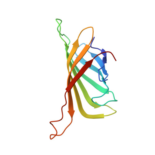
Single particle cryo-EM reconstruction of 52 kDa streptavidin at 3.2 Angstrom resolution.

Fan, X.Wang, J.Zhang, X.Yang, Z.Zhang, J.C.Zhao, L.Peng, H.L.Lei, J.Wang, H.W.

(2019) Nat Commun 10: 2386-2386

  • DOI: https://doi.org/10.1038/s41467-019-10368-w
  • Primary Citation Related Structures: 
    6J6J, 6J6K

  • PubMed Abstract: 

    The fast development of single-particle cryogenic electron microscopy (cryo-EM) has made it more feasible to obtain the 3D structure of well-behaved macromolecules with a molecular weight higher than 300 kDa at ~3 Å resolution. However, it remains a challenge to obtain the high-resolution structures of molecules smaller than 200 kDa using single-particle cryo-EM. In this work, we apply the Cs-corrector-VPP-coupled cryo-EM to study the 52 kDa streptavidin (SA) protein supported on a thin layer of graphene and embedded in vitreous ice. We are able to solve both the apo-SA and biotin-bound SA structures at near-atomic resolution using single-particle cryo-EM. We demonstrate that the method has the potential to determine the structures of molecules as small as 39 kDa.


  • Organizational Affiliation
    • Ministry of Education Key Laboratory of Protein Sciences, Beijing Advanced Innovation Center for Structural Biology, Beijing Frontier Research Center of Biological Structures, School of Life Sciences, Tsinghua University, Beijing, 100084, China.

Macromolecule Content 

  • Total Structure Weight: 50.39 kDa 
  • Atom Count: 3,548 
  • Modeled Residue Count: 476 
  • Deposited Residue Count: 476 
  • Unique protein chains: 1

Macromolecules

Find similar proteins by:|  3D Structure
Entity ID: 1
MoleculeChains  Sequence LengthOrganismDetailsImage
Streptavidin
A, B, C, D
119Streptomyces avidiniiMutation(s): 0 
UniProt
Find proteins for P22629 (Streptomyces avidinii)
Explore P22629 
Go to UniProtKB:  P22629
Entity Groups
Sequence Clusters30% Identity50% Identity70% Identity90% Identity95% Identity100% Identity
UniProt GroupP22629
Sequence Annotations
Expand
Reference Sequence

Experimental Data & Validation

Experimental Data

  • Method: ELECTRON MICROSCOPY
  • Resolution: 3.30 Å
  • Aggregation State: PARTICLE 
  • Reconstruction Method: SINGLE PARTICLE 
EM Software:
TaskSoftware PackageVersion
MODEL REFINEMENTPHENIX
RECONSTRUCTIONRELION

Structure Validation

View Full Validation Report


Entry History 

& Funding Information

Deposition Data


Funding OrganizationLocationGrant Number
Ministry of Science and Technology (China)China2016YFA0501100 to H.W.

Revision History  (Full details and data files)

  • Version 1.0: 2019-05-29
    Type: Initial release
  • Version 1.1: 2019-06-19
    Changes: Data collection, Database references
  • Version 1.2: 2019-11-06
    Changes: Data collection, Other
  • Version 1.3: 2020-08-05
    Changes: Data collection
  • Version 1.4: 2024-03-27
    Changes: Data collection, Database references
  • Version 1.5: 2025-07-02
    Changes: Data collection, Structure summary