6IWJ

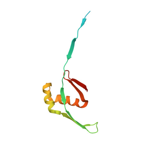
A designed domain swapped dimer


Experimental Data Snapshot

  • Method: SOLUTION NMR
  • Conformers Calculated: 200 
  • Conformers Submitted: 20 
  • Selection Criteria: structures with the lowest energy 

wwPDB Validation   3D Report Full Report


This is version 1.1 of the entry. See complete history


Literature

A five-residue motif for the design of domain swapping in proteins.

Nandwani, N.Surana, P.Negi, H.Mascarenhas, N.M.Udgaonkar, J.B.Das, R.Gosavi, S.

(2019) Nat Commun 10: 452-452

  • DOI: https://doi.org/10.1038/s41467-019-08295-x
  • Primary Citation of Related Structures:  
    5YCT, 5YCU, 5YCW, 6IWJ

  • PubMed Abstract: 
  • Domain swapping is the process by which identical monomeric proteins exchange structural elements to generate dimers/oligomers. Although engineered domain swapping is a compelling strategy for protein assembly, its application has been limited due to the lack of simple and reliable design approaches ...

    Domain swapping is the process by which identical monomeric proteins exchange structural elements to generate dimers/oligomers. Although engineered domain swapping is a compelling strategy for protein assembly, its application has been limited due to the lack of simple and reliable design approaches. Here, we demonstrate that the hydrophobic five-residue 'cystatin motif' (QVVAG) from the domain-swapping protein Stefin B, when engineered into a solvent-exposed, tight surface loop between two β-strands prevents the loop from folding back upon itself, and drives domain swapping in non-domain-swapping proteins. High-resolution structural studies demonstrate that engineering the QVVAG stretch independently into various surface loops of four structurally distinct non-domain-swapping proteins enabled the design of different modes of domain swapping in these proteins, including single, double and open-ended domain swapping. These results suggest that the introduction of the QVVAG motif can be used as a mutational approach for engineering domain swapping in diverse β-hairpin proteins.


    Organizational Affiliation

    National Centre for Biological Sciences, Tata Institute of Fundamental Research, Bengaluru, 560065, India. shachi@ncbs.res.in.


Macromolecules
Find similar proteins by:  (by identity cutoff)  |  3D Structure
Entity ID: 1
MoleculeChainsSequence LengthOrganismDetailsImage
Archeal Protein MK0293
A, B
102Methanopyrus kandleri AV19Mutation(s): 5 
Gene Names: MK0293
UniProt
Find proteins for Q8TYK3 (Methanopyrus kandleri (strain AV19 / DSM 6324 / JCM 9639 / NBRC 100938))
Explore Q8TYK3 
Go to UniProtKB:  Q8TYK3
Entity Groups  
Sequence Clusters30% Identity50% Identity70% Identity90% Identity95% Identity100% Identity
UniProt GroupQ8TYK3
Protein Feature View
Expand
  • Reference Sequence
Experimental Data & Validation

Experimental Data

  • Method: SOLUTION NMR
  • Conformers Calculated: 200 
  • Conformers Submitted: 20 
  • Selection Criteria: structures with the lowest energy 

Structure Validation

View Full Validation Report


Entry History 

Deposition Data

Revision History  (Full details and data files)

  • Version 1.0: 2019-02-13
    Type: Initial release
  • Version 1.1: 2023-06-14
    Changes: Data collection, Database references, Other