6IQG | pdb_00006iqg

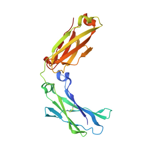
X-ray crystal structure of Fc and peptide complex


Experimental Data Snapshot

  • Method: X-RAY DIFFRACTION
  • Resolution: 3.00 Å
  • R-Value Free: 
    0.273 (Depositor), 0.274 (DCC) 
  • R-Value Work: 
    0.198 (Depositor), 0.203 (DCC) 
  • R-Value Observed: 
    0.202 (Depositor) 

Starting Model: experimental
View more details

wwPDB Validation 3D Report Full Report

Validation slider image for 6IQG

This is version 3.1 of the entry. See complete history. 

Literature

Site-Specific Chemical Conjugation of Antibodies by Using Affinity Peptide for the Development of Therapeutic Antibody Format.

Kishimoto, S., Nakashimada, Y., Yokota, R., Hatanaka, T., Adachi, M., Ito, Y.

(2019) Bioconjug Chem 30: 698-702

  • DOI: https://doi.org/10.1021/acs.bioconjchem.8b00865
  • Primary Citation Related Structures: 
    6IQG, 6IQH

  • PubMed Abstract: 

    Artificially modified IgG molecules are increasingly utilized in industrial and clinical applications. In the present study, the method of chemical conjugation by affinity peptide (CCAP) for site-specific chemical modification has been developed by using a peptide that bound with high affinity to human IgG-Fc. This method enabled a rapid modification of a specific residue (Lys248 on Fc) in a one-step reaction under mild condition to form a stable amide bond between the peptide and Fc. The monovalent peptide-IgG conjugate not only maintained complete antigen binding but also bound to Fc receptors (FcRn, FcγRI, and FcγRIIIa), indicating that it is a suitable conjugate form that can be further developed into highly functional antibody therapeutics. CCAP was applied for the preparation of an antibody-drug conjugate and a bispecific antibody to demonstrate the usefulness of this method.


  • Organizational Affiliation: 
    • Graduate School of Science and Engineering , Kagoshima University , Kagoshima 890-0065 , Japan.

Macromolecule Content 

  • Total Structure Weight: 54.5 kDa 
  • Atom Count: 3,794 
  • Modeled Residue Count: 442 
  • Deposited Residue Count: 456 
  • Unique protein chains: 2

Macromolecules

Find similar proteins by:|  3D Structure
Entity ID: 1
MoleculeChains  Sequence LengthOrganismDetailsImage
Immunoglobulin gamma-1 heavy chain
A, B
210Homo sapiensMutation(s): 0 
UniProt
Find proteins for P0DOX5 (Homo sapiens)
Explore P0DOX5 
Go to UniProtKB:  P0DOX5
Entity Groups
Sequence Clusters30% Identity50% Identity70% Identity90% Identity95% Identity100% Identity
UniProt GroupP0DOX5
Glycosylation
Glycosylation Sites: 1
Sequence Annotations
Expand
Reference Sequence
Find similar proteins by:  Sequence   |   3D Structure  
Entity ID: 2
MoleculeChains  Sequence LengthOrganismDetailsImage
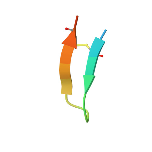
18-mer peptide G(HCS)DCAYHRGELVWCT(HCS)H(NH2)
C, D
18synthetic constructMutation(s): 0 
Entity Groups
Sequence Clusters30% Identity50% Identity70% Identity90% Identity95% Identity100% Identity
Sequence Annotations
Expand
Reference Sequence

Oligosaccharides

Help  
Entity ID: 3
MoleculeChains Length2D Diagram GlycosylationD Interactions
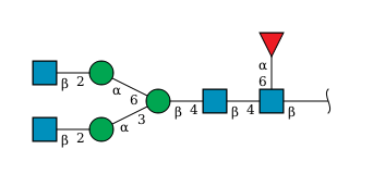
2-acetamido-2-deoxy-beta-D-glucopyranose-(1-2)-alpha-D-mannopyranose-(1-3)-[2-acetamido-2-deoxy-beta-D-glucopyranose-(1-2)-alpha-D-mannopyranose-(1-6)]beta-D-mannopyranose-(1-4)-2-acetamido-2-deoxy-beta-D-glucopyranose-(1-4)-[alpha-L-fucopyranose-(1-6)]2-acetamido-2-deoxy-beta-D-glucopyranose
E, F
8N-Glycosylation
Glycosylation Resources
GlyTouCan: G80858MF
GlyCosmos: G80858MF
GlyGen: G80858MF

Small Molecules

Ligands 1 Unique
IDChains Name / Formula / InChI Key2D Diagram3D Interactions
CL

Query on CL


Download:Ideal Coordinates CCD File
G [auth A]CHLORIDE ION
Cl
VEXZGXHMUGYJMC-UHFFFAOYSA-M
Modified Residues  1 Unique
IDChains TypeFormula2D DiagramParent
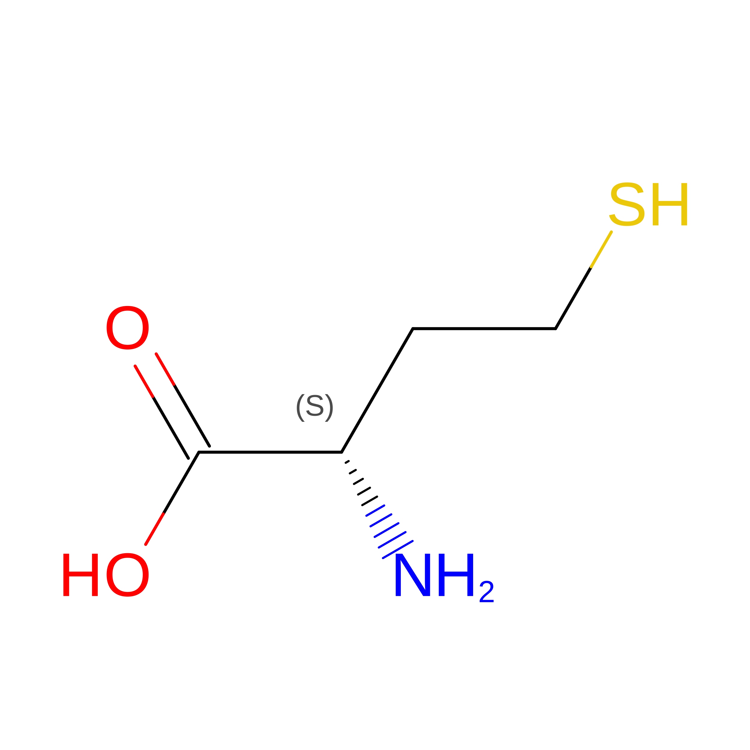
HCS
Query on HCS
C, D
L-PEPTIDE LINKINGC4 H9 N O2 S

--

Experimental Data & Validation

Experimental Data

  • Method: X-RAY DIFFRACTION
  • Resolution: 3.00 Å
  • R-Value Free:  0.273 (Depositor), 0.274 (DCC) 
  • R-Value Work:  0.198 (Depositor), 0.203 (DCC) 
  • R-Value Observed: 0.202 (Depositor) 
Space Group: P 1 21 1
Unit Cell:
Length ( Å )Angle ( ˚ )
a = 66.062α = 90
b = 60.507β = 101.25
c = 69.526γ = 90
Software Package:
Software NamePurpose
PHENIXrefinement
HKL-2000data reduction
SCALEPACKdata scaling
PHENIXphasing

Structure Validation

View Full Validation Report


Entry History 

Deposition Data

Revision History  (Full details and data files)

  • Version 1.0: 2019-02-13
    Type: Initial release
  • Version 1.1: 2019-03-13
    Changes: Data collection, Database references
  • Version 1.2: 2019-04-03
    Changes: Data collection, Database references
  • Version 2.0: 2020-07-29
    Type: Remediation
    Reason: Carbohydrate remediation
    Changes: Advisory, Atomic model, Data collection, Derived calculations, Structure summary
  • Version 2.1: 2023-11-22
    Changes: Data collection, Database references, Derived calculations, Refinement description, Structure summary
  • Version 3.0: 2024-03-13
    Changes: Atomic model, Data collection, Structure summary
  • Version 3.1: 2024-11-13
    Changes: Structure summary