6IQG | pdb_00006iqg

X-ray crystal structure of Fc and peptide complex


Experimental Data Snapshot

  • Method: X-RAY DIFFRACTION
  • Resolution: 3.00 Å
  • R-Value Free: 
    0.273 (Depositor), 0.274 (DCC) 
  • R-Value Work: 
    0.198 (Depositor), 0.203 (DCC) 
  • R-Value Observed: 
    0.202 (Depositor) 

Starting Model: experimental
View more details

wwPDB Validation 3D Report Full Report

Validation slider image for 6IQG

This is version 3.1 of the entry. See complete history

Literature

Site-Specific Chemical Conjugation of Antibodies by Using Affinity Peptide for the Development of Therapeutic Antibody Format.

Kishimoto, S.Nakashimada, Y.Yokota, R.Hatanaka, T.Adachi, M.Ito, Y.

(2019) Bioconjug Chem 30: 698-702

  • DOI: https://doi.org/10.1021/acs.bioconjchem.8b00865
  • Primary Citation Related Structures: 
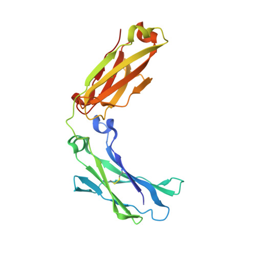
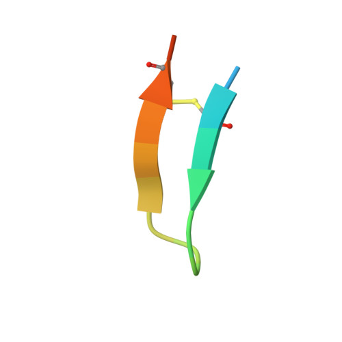
    6IQG, 6IQH

  • PubMed Abstract: 

    Artificially modified IgG molecules are increasingly utilized in industrial and clinical applications. In the present study, the method of chemical conjugation by affinity peptide (CCAP) for site-specific chemical modification has been developed by using a peptide that bound with high affinity to human IgG-Fc. This method enabled a rapid modification of a specific residue (Lys248 on Fc) in a one-step reaction under mild condition to form a stable amide bond between the peptide and Fc. The monovalent peptide-IgG conjugate not only maintained complete antigen binding but also bound to Fc receptors (FcRn, FcγRI, and FcγRIIIa), indicating that it is a suitable conjugate form that can be further developed into highly functional antibody therapeutics. CCAP was applied for the preparation of an antibody-drug conjugate and a bispecific antibody to demonstrate the usefulness of this method.


  • Organizational Affiliation
    • Graduate School of Science and Engineering , Kagoshima University , Kagoshima 890-0065 , Japan.

Macromolecule Content 

  • Total Structure Weight: 54.5 kDa 
  • Atom Count: 3,794 
  • Modeled Residue Count: 442 
  • Deposited Residue Count: 456 
  • Unique protein chains: 2

Macromolecules

Find similar proteins by:|  3D Structure
Entity ID: 1
MoleculeChains  Sequence LengthOrganismDetailsImage
Immunoglobulin gamma-1 heavy chain
A, B
210Homo sapiensMutation(s): 0 
UniProt
Find proteins for P0DOX5 (Homo sapiens)
Explore P0DOX5 
Go to UniProtKB:  P0DOX5
Entity Groups
Sequence Clusters30% Identity50% Identity70% Identity90% Identity95% Identity100% Identity
UniProt GroupP0DOX5
Glycosylation
Glycosylation Sites: 1
Sequence Annotations
Expand
Reference Sequence
Find similar proteins by:  Sequence   |   3D Structure  
Entity ID: 2
MoleculeChains  Sequence LengthOrganismDetailsImage
18-mer peptide G(HCS)DCAYHRGELVWCT(HCS)H(NH2)
C, D
18synthetic constructMutation(s): 0 

Oligosaccharides

Help  
Entity ID: 3
MoleculeChains Length2D Diagram GlycosylationD Interactions
2-acetamido-2-deoxy-beta-D-glucopyranose-(1-2)-alpha-D-mannopyranose-(1-3)-[2-acetamido-2-deoxy-beta-D-glucopyranose-(1-2)-alpha-D-mannopyranose-(1-6)]beta-D-mannopyranose-(1-4)-2-acetamido-2-deoxy-beta-D-glucopyranose-(1-4)-[alpha-L-fucopyranose-(1-6)]2-acetamido-2-deoxy-beta-D-glucopyranose
E, F
8N-Glycosylation
Glycosylation Resources
GlyTouCan: G80858MF
GlyCosmos: G80858MF
GlyGen: G80858MF

Small Molecules

Ligands 1 Unique
IDChains Name / Formula / InChI Key2D Diagram3D Interactions
CL

Query on CL


Download:Ideal Coordinates CCD File
G [auth A]CHLORIDE ION
Cl
VEXZGXHMUGYJMC-UHFFFAOYSA-M

Experimental Data & Validation

Experimental Data

  • Method: X-RAY DIFFRACTION
  • Resolution: 3.00 Å
  • R-Value Free:  0.273 (Depositor), 0.274 (DCC) 
  • R-Value Work:  0.198 (Depositor), 0.203 (DCC) 
  • R-Value Observed: 0.202 (Depositor) 
Space Group: P 1 21 1
Unit Cell:
Length ( Å )Angle ( ˚ )
a = 66.062α = 90
b = 60.507β = 101.25
c = 69.526γ = 90
Software Package:
Software NamePurpose
PHENIXrefinement
HKL-2000data reduction
SCALEPACKdata scaling
PHENIXphasing

Structure Validation

View Full Validation Report


Entry History 

Deposition Data

Revision History  (Full details and data files)

  • Version 1.0: 2019-02-13
    Type: Initial release
  • Version 1.1: 2019-03-13
    Changes: Data collection, Database references
  • Version 1.2: 2019-04-03
    Changes: Data collection, Database references
  • Version 2.0: 2020-07-29
    Type: Remediation
    Reason: Carbohydrate remediation
    Changes: Advisory, Atomic model, Data collection, Derived calculations, Structure summary
  • Version 2.1: 2023-11-22
    Changes: Data collection, Database references, Derived calculations, Refinement description, Structure summary
  • Version 3.0: 2024-03-13
    Changes: Atomic model, Data collection, Structure summary
  • Version 3.1: 2024-11-13
    Changes: Structure summary