6IF1 | pdb_00006if1

Crystal structure of Ube2K and K48-linked di-ubiquitin complex


Experimental Data Snapshot

  • Method: X-RAY DIFFRACTION
  • Resolution: 2.47 Å
  • R-Value Free: 
    0.236 (Depositor), 0.236 (DCC) 
  • R-Value Work: 
    0.215 (Depositor), 0.215 (DCC) 
  • R-Value Observed: 
    0.216 (Depositor) 

Starting Model: experimental
View more details

wwPDB Validation 3D Report Full Report

Validation slider image for 6IF1

This is version 1.1 of the entry. See complete history

Literature

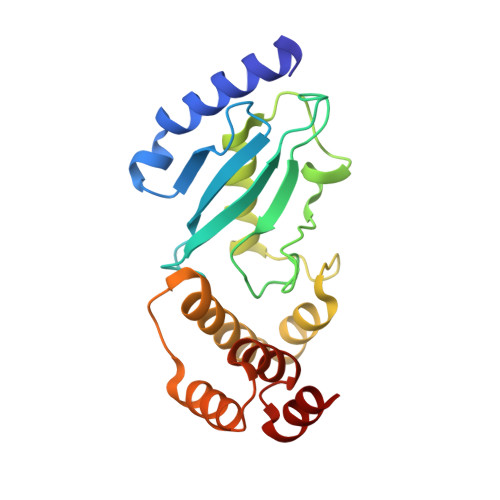
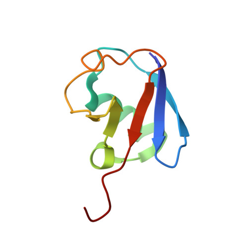
Crystal structure of the Ube2K/E2-25K and K48-linked di-ubiquitin complex provides structural insight into the mechanism of K48-specific ubiquitin chain synthesis.

Lee, J.G.Youn, H.S.Kang, J.Y.Park, S.Y.Kidera, A.Yoo, Y.J.Eom, S.H.

(2018) Biochem Biophys Res Commun 506: 102-107

  • DOI: https://doi.org/10.1016/j.bbrc.2018.10.067
  • Primary Citation Related Structures: 
    6IF1

  • PubMed Abstract: 

    Ubiquitin-conjugating enzymes (E2) form thioester bonds with ubiquitin (Ub), which are subsequently transferred to target proteins for cellular progress. Ube2K/E2-25K (a class II E2 enzyme) contains a C-terminal ubiquitin-associated (UBA) domain that has been suggested to control ubiquitin recognition, dimerization, or poly-ubiquitin chain formation. Ube2K is a special E2 because it synthesizes K48-linked poly-ubiquitin chains without E3 ubiquitin ligase. We found that a novel interaction between the acceptor di-Ub (Ub 2 ) and the auxiliary Ube2K promotes the discharging reaction and production of tri-Ub (Ub 3 ), probably by guiding and positioning the K48 (in the distal Ub) of the acceptor Ub 2 in the active site. We also determined the crystal structure of Ube2K-Ub 2 at 2.47 Å resolution. Based on our structural and biochemical data, we proposed a structural model of Ub 3 synthesis by Ube2K without E3.


  • Organizational Affiliation
    • School of Life Sciences, Gwangju Institute of Science and Technology, Gwangju, 61005, Republic of Korea; Steitz Center for Structural Biology, Gwangju Institute of Science and Technology, Gwangju, 61005, Republic of Korea.

Macromolecule Content 

  • Total Structure Weight: 61.79 kDa 
  • Atom Count: 4,366 
  • Modeled Residue Count: 550 
  • Deposited Residue Count: 550 
  • Unique protein chains: 2

Macromolecules

Find similar proteins by:|  3D Structure
Entity ID: 1
MoleculeChains  Sequence LengthOrganismDetailsImage
Ubiquitin-conjugating enzyme E2 K
A, B
199Homo sapiensMutation(s): 0 
Gene Names: UBE2KHIP2LIG
EC: 2.3.2.23
UniProt & NIH Common Fund Data Resources
Find proteins for P61086 (Homo sapiens)
Explore P61086 
Go to UniProtKB:  P61086
PHAROS:  P61086
GTEx:  ENSG00000078140 
Entity Groups
Sequence Clusters30% Identity50% Identity70% Identity90% Identity95% Identity100% Identity
UniProt GroupP61086
Sequence Annotations
Expand
Reference Sequence
Find similar proteins by:|  3D Structure
Entity ID: 2
MoleculeChains  Sequence LengthOrganismDetailsImage
Ubiquitin
C, D
76Homo sapiensMutation(s): 0 
Gene Names: UBB
UniProt & NIH Common Fund Data Resources
Find proteins for P0CG47 (Homo sapiens)
Explore P0CG47 
Go to UniProtKB:  P0CG47
PHAROS:  P0CG47
GTEx:  ENSG00000170315 
Entity Groups
Sequence Clusters30% Identity50% Identity70% Identity90% Identity95% Identity100% Identity
UniProt GroupP0CG47
Sequence Annotations
Expand
Reference Sequence

Experimental Data & Validation

Experimental Data

  • Method: X-RAY DIFFRACTION
  • Resolution: 2.47 Å
  • R-Value Free:  0.236 (Depositor), 0.236 (DCC) 
  • R-Value Work:  0.215 (Depositor), 0.215 (DCC) 
  • R-Value Observed: 0.216 (Depositor) 
Space Group: P 1 21 1
Unit Cell:
Length ( Å )Angle ( ˚ )
a = 41.559α = 90
b = 38.792β = 90.1
c = 190.321γ = 90
Software Package:
Software NamePurpose
PHENIXrefinement
HKL-2000data reduction
HKL-2000data scaling
PHASERphasing

Structure Validation

View Full Validation Report


Entry History 

Deposition Data

Revision History  (Full details and data files)

  • Version 1.0: 2018-11-21
    Type: Initial release
  • Version 1.1: 2023-11-22
    Changes: Data collection, Database references, Refinement description