6I0X

Porphyromonas gingivalis peptidylarginine deminase (PPAD) mutant G231N/E232T/N235D in complex with Cl-amidine.


Experimental Data Snapshot

  • Method: X-RAY DIFFRACTION
  • Resolution: 1.60 Å
  • R-Value Free: 0.193 
  • R-Value Work: 0.180 
  • R-Value Observed: 0.180 

wwPDB Validation   3D Report Full Report


Ligand Structure Quality Assessment 


This is version 1.3 of the entry. See complete history


Literature

Structure, function, and inhibition of a genomic/clinical variant of Porphyromonas gingivalis peptidylarginine deiminase.

Bereta, G.Goulas, T.Madej, M.Bielecka, E.Sola, M.Potempa, J.Xavier Gomis-Ruth, F.

(2019) Protein Sci 28: 478-486

  • DOI: https://doi.org/10.1002/pro.3571
  • Primary Citation of Related Structures:  
    6I0X

  • PubMed Abstract: 

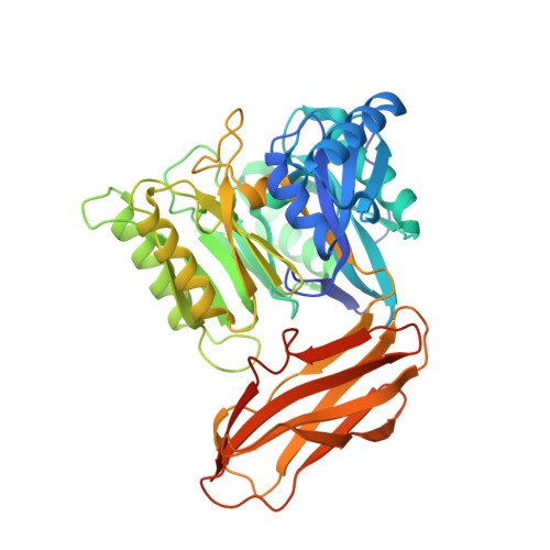
    Citrullination is an essential post-translational modification in which the guanidinium group of protein and peptide arginines is deiminated by peptidylarginine deiminases (PADs). When deregulated, excessive citrullination leads to inflammation as in severe periodontal disease (PD) and rheumatoid arthritis (RA). Porphyromonas gingivalis is the major periodontopathogenic causative agent of PD and also an etiological agent of RA. It secretes a PAD, termed Porphyromonas PAD (PPAD), which is a virulence factor that causes aberrant citrullination. Analysis of P. gingivalis genomes of laboratory strains and clinical isolates unveiled a PPAD variant (PPAD-T2), which showed three amino-acid substitutions directly preceding catalytic Residue H 236 (G 231 N/E 232 T/N 235 D) when compared with PPAD from the reference strain (PPAD-T1). Mutation of these positions in the reference strain resulted in twofold higher cell-associated citrullinating activity. Similar to PPAD-T1, recombinant PPAD-T2 citrullinated arginines at the C-termini of general peptidic substrates but not within peptides. Catalytically, PPAD-T2 showed weaker substrate binding but higher turnover rates than PPAD-T1. In contrast, no differences were found in thermal stability. The 1.6 Å-resolution X-ray crystal structure of PPAD-T2 in complex with the general human PAD inhibitor, Cl-amidine, revealed that the inhibitor moiety is tightly bound and that mutations localize to a loop engaged in substrate/inhibitor binding. In particular, mutation G 231 N caused a slight structural rearrangement, which probably originated the higher substrate turnover observed. The present data compare two natural PPAD variants and will set the pace for the design of specific inhibitors against P. gingivalis-caused PD.


  • Organizational Affiliation

    Department of Microbiology, Faculty of Biochemistry, Biophysics and Biotechnology, Jagiellonian University, Gronostajowa 7, PL-30-387, Kraków, Poland.


Macromolecules
Find similar proteins by:  (by identity cutoff)  |  3D Structure
Entity ID: 1
MoleculeChains Sequence LengthOrganismDetailsImage
Peptidylarginine deiminase
A, B
438Porphyromonas gingivalis W83Mutation(s): 3 
Gene Names: PG_1424
EC: 3.5.3
UniProt
Find proteins for Q9RQJ2 (Porphyromonas gingivalis (strain ATCC BAA-308 / W83))
Explore Q9RQJ2 
Go to UniProtKB:  Q9RQJ2
Entity Groups  
Sequence Clusters30% Identity50% Identity70% Identity90% Identity95% Identity100% Identity
UniProt GroupQ9RQJ2
Sequence Annotations
Expand
  • Reference Sequence
Small Molecules
Ligands 3 Unique
IDChains Name / Formula / InChI Key2D Diagram3D Interactions
BFB
Query on BFB

Download Ideal Coordinates CCD File 
D [auth A],
J [auth B]
N-[(1S)-1-(AMINOCARBONYL)-4-(ETHANIMIDOYLAMINO)BUTYL]BENZAMIDE
C14 H20 N4 O2
LWFFSSMDFWZNNW-LBPRGKRZSA-N
GOL
Query on GOL

Download Ideal Coordinates CCD File 
F [auth A],
G [auth A],
H [auth A],
K [auth B],
L [auth B]
GLYCEROL
C3 H8 O3
PEDCQBHIVMGVHV-UHFFFAOYSA-N
NA
Query on NA

Download Ideal Coordinates CCD File 
C [auth A],
E [auth A],
I [auth B]
SODIUM ION
Na
FKNQFGJONOIPTF-UHFFFAOYSA-N
Experimental Data & Validation

Experimental Data

  • Method: X-RAY DIFFRACTION
  • Resolution: 1.60 Å
  • R-Value Free: 0.193 
  • R-Value Work: 0.180 
  • R-Value Observed: 0.180 
  • Space Group: C 1 2 1
Unit Cell:
Length ( Å )Angle ( ˚ )
a = 123.84α = 90
b = 71.87β = 95.86
c = 104.72γ = 90
Software Package:
Software NamePurpose
BUSTERrefinement
XDSdata reduction
XSCALEdata scaling
PHASERphasing

Structure Validation

View Full Validation Report


Ligand Structure Quality Assessment 


Entry History 

Deposition Data

Revision History  (Full details and data files)

  • Version 1.0: 2019-01-23
    Type: Initial release
  • Version 1.1: 2019-01-30
    Changes: Data collection, Database references
  • Version 1.2: 2019-02-20
    Changes: Data collection, Database references
  • Version 1.3: 2024-01-24
    Changes: Data collection, Database references, Derived calculations, Refinement description