6I0V | pdb_00006i0v

Crystal structure of DmTailor in complex with CACAGU RNA


Experimental Data Snapshot

  • Method: X-RAY DIFFRACTION
  • Resolution: 1.85 Å
  • R-Value Free: 
    0.226 (Depositor), 0.229 (DCC) 
  • R-Value Work: 
    0.195 (Depositor), 0.198 (DCC) 
  • R-Value Observed: 
    0.196 (Depositor) 

Starting Model: experimental
View more details

wwPDB Validation 3D Report Full Report

Validation slider image for 6I0V

This is version 1.2 of the entry. See complete history

Literature

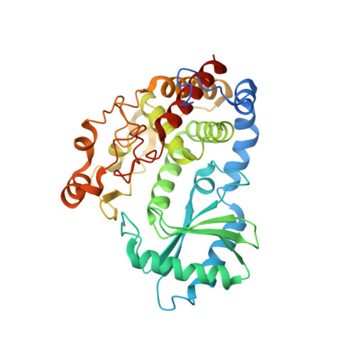
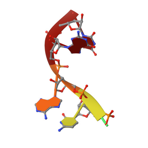
Structural basis for acceptor RNA substrate selectivity of the 3' terminal uridylyl transferase Tailor.

Kroupova, A.Ivascu, A.Reimao-Pinto, M.M.Ameres, S.L.Jinek, M.

(2019) Nucleic Acids Res 47: 1030-1042

  • DOI: https://doi.org/10.1093/nar/gky1164
  • Primary Citation Related Structures: 
    6I0S, 6I0T, 6I0U, 6I0V

  • PubMed Abstract: 

    Non-templated 3'-uridylation of RNAs has emerged as an important mechanism for regulating the processing, stability and biological function of eukaryotic transcripts. In Drosophila, oligouridine tailing by the terminal uridylyl transferase (TUTase) Tailor of numerous RNAs induces their degradation by the exonuclease Dis3L2, which serves functional roles in RNA surveillance and mirtron RNA biogenesis. Tailor preferentially uridylates RNAs terminating in guanosine or uridine nucleotides but the structural basis underpinning its RNA substrate selectivity is unknown. Here, we report crystal structures of Tailor bound to a donor substrate analog or mono- and oligouridylated RNA products. These structures reveal specific amino acid residues involved in donor and acceptor substrate recognition, and complementary biochemical assays confirm the critical role of an active site arginine in conferring selectivity toward 3'-guanosine terminated RNAs. Notably, conservation of these active site features suggests that other eukaryotic TUTases, including mammalian TUT4 and TUT7, might exhibit similar, hitherto unknown, substrate selectivity. Together, these studies provide critical insights into the specificity of 3'-uridylation in eukaryotic post-transcriptional gene regulation.


  • Organizational Affiliation
    • Department of Biochemistry, University of Zurich, Zurich 8057, Switzerland.

Macromolecule Content 

  • Total Structure Weight: 46.18 kDa 
  • Atom Count: 3,018 
  • Modeled Residue Count: 351 
  • Deposited Residue Count: 390 
  • Unique protein chains: 1
  • Unique nucleic acid chains: 1

Macromolecules


Find similar proteins by:|  3D Structure
Entity ID: 1
MoleculeChains  Sequence LengthOrganismDetailsImage
Terminal uridylyltransferase Tailor384Drosophila melanogasterMutation(s): 0 
Gene Names: TailorCG1091
EC: 2.7.7.52
UniProt
Find proteins for Q9VI58 (Drosophila melanogaster)
Explore Q9VI58 
Go to UniProtKB:  Q9VI58
Entity Groups
Sequence Clusters30% Identity50% Identity70% Identity90% Identity95% Identity100% Identity
UniProt GroupQ9VI58
Sequence Annotations
Expand
Reference Sequence
Find similar nucleic acids by:  Sequence
Entity ID: 2
MoleculeChains LengthOrganismImage
RNA (5'-R(*CP*AP*CP*AP*GP*U)-3')6Drosophila melanogaster
Sequence Annotations
Expand
Reference Sequence

Experimental Data & Validation

Experimental Data

  • Method: X-RAY DIFFRACTION
  • Resolution: 1.85 Å
  • R-Value Free:  0.226 (Depositor), 0.229 (DCC) 
  • R-Value Work:  0.195 (Depositor), 0.198 (DCC) 
  • R-Value Observed: 0.196 (Depositor) 
Space Group: P 31 2 1
Unit Cell:
Length ( Å )Angle ( ˚ )
a = 60.077α = 90
b = 60.077β = 90
c = 162.293γ = 120
Software Package:
Software NamePurpose
PHENIXrefinement
XDSdata reduction
Cootmodel building
PHASERphasing
XSCALEdata scaling

Structure Validation

View Full Validation Report


Entry History 

Deposition Data

Revision History  (Full details and data files)

  • Version 1.0: 2018-12-05
    Type: Initial release
  • Version 1.1: 2019-02-06
    Changes: Data collection, Database references
  • Version 1.2: 2024-01-24
    Changes: Data collection, Database references, Derived calculations, Refinement description