Structural determinants increasing flexibility confer cold adaptation in psychrophilic phosphoglycerate kinase.
Mandelman, D., Ballut, L., Wolff, D.A., Feller, G., Gerday, C., Haser, R., Aghajari, N.(2019) Extremophiles 23: 495-506
- PubMed: 31147836 Search on PubMed
- DOI: https://doi.org/10.1007/s00792-019-01102-x
- Primary Citation Related Structures:
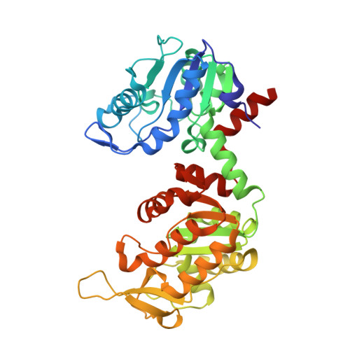
6HXE, 6I06 - PubMed Abstract:
Crystal structures of phosphoglycerate kinase (PGK) from the psychrophile Pseudomonas sp. TACII 18 have been determined at high resolution by X-ray crystallography methods and compared with mesophilic, thermophilic and hyperthermophilic counterparts. PGK is a two-domain enzyme undergoing large domain movements to catalyze the production of ATP from 1,3-biphosphoglycerate and ADP. Whereas the conformational dynamics sustaining the catalytic mechanism of this hinge-bending enzyme now seems rather clear, the determinants which underlie high catalytic efficiency at low temperatures of this psychrophilic PGK were unknown. The comparison of the three-dimensional structures shows that multiple (global and local) specific adaptations have been brought about by this enzyme. Together, these reside in an overall increased flexibility of the cold-adapted PGK thereby allowing a better accessibility to the active site, but also a potentially more disordered transition state of the psychrophilic enzyme, due to the destabilization of some catalytic residues.
- Biocrystallography and Structural Biology of Therapeutic Targets, Molecular Microbiology and Structural Biochemistry, UMR 5086, CNRS, University of Lyon 1, 7 passage du Vercors, 69367, Lyon Cedex 07, France.
Organizational Affiliation: