6HXA

AntI from P. luminescens catalyses terminal polyketide shortening in the biosynthesis of anthraquinones


Experimental Data Snapshot

  • Method: X-RAY DIFFRACTION
  • Resolution: 1.85 Å
  • R-Value Free: 0.204 
  • R-Value Work: 0.168 
  • R-Value Observed: 0.170 

wwPDB Validation   3D Report Full Report


This is version 1.0 of the entry. See complete history


Literature

Molecular mechanism of polyketide shortening in anthraquinone biosynthesis ofPhotorhabdus luminescens.

Zhou, Q.Brauer, A.Adihou, H.Schmalhofer, M.Saura, P.Grammbitter, G.L.C.Kaila, V.R.I.Groll, M.Bode, H.B.

(2019) Chem Sci 10: 6341-6349

  • DOI: https://doi.org/10.1039/c9sc00749k
  • Primary Citation of Related Structures:  
    6HXA

  • PubMed Abstract: 

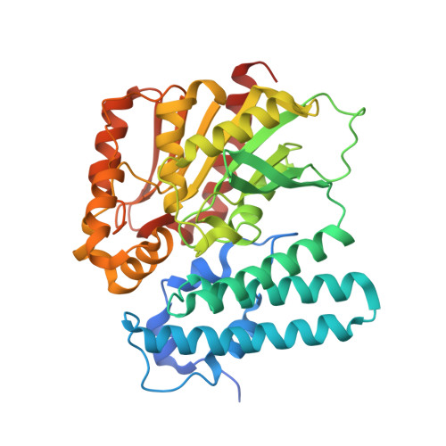
    Anthraquinones, a widely distributed class of aromatic natural products, are produced by a type II polyketide synthase system in the Gram-negative bacterium Photorhabdus luminescens . Heterologous expression of the antABCDEFGHI anthraquinone biosynthetic gene cluster in Escherichia coli identified AntI as an unusual lyase, catalysing terminal polyketide shortening prior to formation of the third aromatic ring. Functional in vitro and in vivo analysis of AntI using X-ray crystallography, structure-based mutagenesis, and molecular simulations revealed that AntI converts a defined octaketide to the tricyclic anthraquinone ring via retro-Claisen and Dieckmann reactions. Thus, AntI catalyses a so far unobserved multistep reaction in this PKS system.


  • Organizational Affiliation

    Molekulare Biotechnologie , Fachbereich Biowissenschaften , Buchmann Institute for Molecular Life Sciences (BMLS) , Goethe Universität Frankfurt , Max-von-Laue-Str. 15, Max-von-Laue-Str. 9 , 60438 Frankfurt am Main , Germany . Email: h.bode@bio.uni-frankfurt.de.


Macromolecules
Find similar proteins by:  (by identity cutoff)  |  3D Structure
Entity ID: 1
MoleculeChains Sequence LengthOrganismDetailsImage
AntI399Photorhabdus luminescensMutation(s): 0 
Gene Names: plu4186
UniProt
Find proteins for Q7MZT8 (Photorhabdus laumondii subsp. laumondii (strain DSM 15139 / CIP 105565 / TT01))
Explore Q7MZT8 
Go to UniProtKB:  Q7MZT8
Entity Groups  
Sequence Clusters30% Identity50% Identity70% Identity90% Identity95% Identity100% Identity
UniProt GroupQ7MZT8
Sequence Annotations
Expand
  • Reference Sequence
Small Molecules
Ligands 2 Unique
IDChains Name / Formula / InChI Key2D Diagram3D Interactions
PG4
Query on PG4

Download Ideal Coordinates CCD File 
C [auth A]TETRAETHYLENE GLYCOL
C8 H18 O5
UWHCKJMYHZGTIT-UHFFFAOYSA-N
PGE
Query on PGE

Download Ideal Coordinates CCD File 
B [auth A]TRIETHYLENE GLYCOL
C6 H14 O4
ZIBGPFATKBEMQZ-UHFFFAOYSA-N
Experimental Data & Validation

Experimental Data

  • Method: X-RAY DIFFRACTION
  • Resolution: 1.85 Å
  • R-Value Free: 0.204 
  • R-Value Work: 0.168 
  • R-Value Observed: 0.170 
  • Space Group: C 2 2 21
Unit Cell:
Length ( Å )Angle ( ˚ )
a = 54.11α = 90
b = 154.34β = 90
c = 91.24γ = 90
Software Package:
Software NamePurpose
REFMACrefinement
XDSdata reduction
XSCALEdata scaling
SHARPphasing

Structure Validation

View Full Validation Report


Entry History & Funding Information

Deposition Data


Funding OrganizationLocationGrant Number
German Research FoundationGermanySFB749

Revision History  (Full details and data files)

  • Version 1.0: 2019-08-07
    Type: Initial release