6HK3

Crystal structure of GSK-3B in complex with pyrazine inhibitor C44


Experimental Data Snapshot

  • Method: X-RAY DIFFRACTION
  • Resolution: 2.35 Å
  • R-Value Free: 0.234 
  • R-Value Work: 0.204 
  • R-Value Observed: 0.206 

wwPDB Validation   3D Report Full Report


Ligand Structure Quality Assessment 


This is version 1.2 of the entry. See complete history


Literature

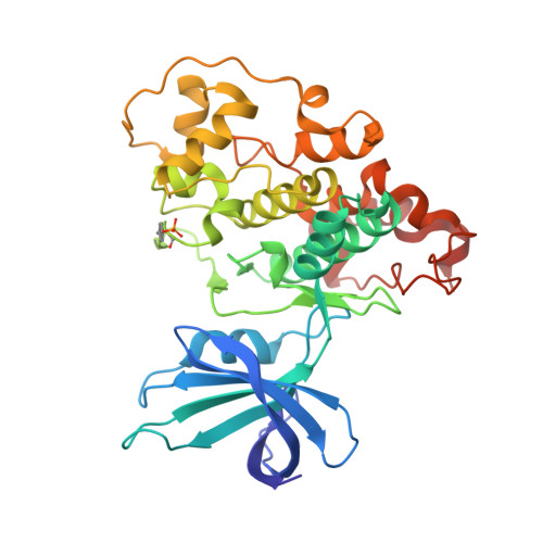
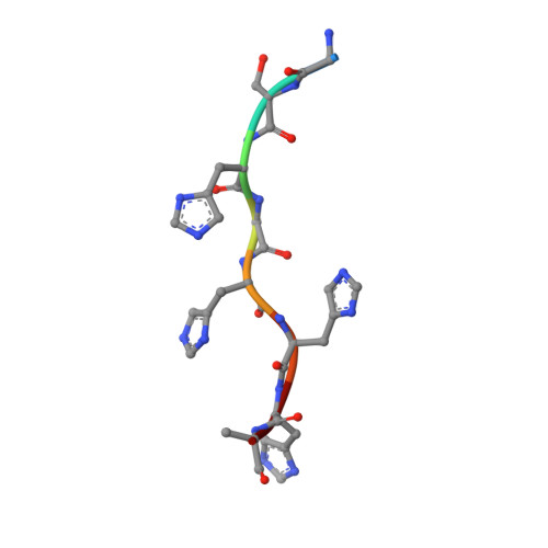
Investigating Drug-Target Residence Time in Kinases through Enhanced Sampling Simulations.

Gobbo, D.Piretti, V.Di Martino, R.M.C.Tripathi, S.K.Giabbai, B.Storici, P.Demitri, N.Girotto, S.Decherchi, S.Cavalli, A.

(2019) J Chem Theory Comput 15: 4646-4659

  • DOI: https://doi.org/10.1021/acs.jctc.9b00104
  • Primary Citation of Related Structures:  
    6HK3, 6HK4, 6HK7

  • PubMed Abstract: 

    It is widely accepted that drug-target association and dissociation rates directly affect drug efficacy and safety. To rationally optimize drug binding kinetics, one must know the atomic arrangement of the protein-ligand complex during the binding/unbinding process in order to detect stable and metastable states. Whereas experimental approaches can determine kinetic constants with fairly good accuracy, computational approaches based on molecular dynamics (MD) simulations can deliver the atomistic details of the unbinding process. Furthermore, they can also be utilized prospectively to predict residence time (i.e., the inverse of unbinding kinetics constant, k off ) with an acceptable level of accuracy. Here, we report a novel method based on adiabatic bias MD with an electrostatics-like collective variable (dubbed elABMD) for sampling protein-ligand dissociation events in two kinases. elABMD correctly ranked a ligand series on glucokinase, in agreement with experimental data and previous calculations. Subsequently, we applied the new method prospectively to a congeneric series of GSK-3β inhibitors. For this series, new crystal structures were generated and the residence time was experimentally measured with surface plasmon resonance (SPR). There was good agreement between computational predictions and experimental measures, suggesting that elABMD is an innovative and efficient tool for calculating residence times.


  • Organizational Affiliation

    Computational and Chemical Biology , Fondazione Istituto Italiano di Tecnologia , via Morego 30 , 16163 Genova , Italy.


Macromolecules
Find similar proteins by:  (by identity cutoff)  |  3D Structure
Entity ID: 1
MoleculeChains Sequence LengthOrganismDetailsImage
Glycogen synthase kinase-3 beta
A, B
350Homo sapiensMutation(s): 0 
Gene Names: GSK3B
EC: 2.7.11.26 (PDB Primary Data), 2.7.11.1 (PDB Primary Data)
UniProt & NIH Common Fund Data Resources
Find proteins for P49841 (Homo sapiens)
Explore P49841 
Go to UniProtKB:  P49841
PHAROS:  P49841
GTEx:  ENSG00000082701 
Entity Groups  
Sequence Clusters30% Identity50% Identity70% Identity90% Identity95% Identity100% Identity
UniProt GroupP49841
Sequence Annotations
Expand
  • Reference Sequence

Find similar proteins by:  Sequence   |   3D Structure  

Entity ID: 2
MoleculeChains Sequence LengthOrganismDetailsImage
GLY-SER-HIS-GLY-HIS-HIS-HIS-HISC [auth N]8Homo sapiensMutation(s): 0 
Sequence Annotations
Expand
  • Reference Sequence
Small Molecules
Ligands 5 Unique
IDChains Name / Formula / InChI Key2D Diagram3D Interactions
G8B (Subject of Investigation/LOI)
Query on G8B

Download Ideal Coordinates CCD File 
D [auth A],
K [auth B]
3-azanyl-~{N}-(2-methoxyphenyl)-6-[4-(4-methylpiperazin-1-yl)sulfonylphenyl]pyrazine-2-carboxamide
C23 H26 N6 O4 S
VUQAIGCULONGGN-UHFFFAOYSA-N
MLI
Query on MLI

Download Ideal Coordinates CCD File 
E [auth A],
L [auth B]
MALONATE ION
C3 H2 O4
OFOBLEOULBTSOW-UHFFFAOYSA-L
GOL
Query on GOL

Download Ideal Coordinates CCD File 
F [auth A],
G [auth A],
H [auth A],
M [auth B]
GLYCEROL
C3 H8 O3
PEDCQBHIVMGVHV-UHFFFAOYSA-N
DMS
Query on DMS

Download Ideal Coordinates CCD File 
J [auth A],
O [auth B]
DIMETHYL SULFOXIDE
C2 H6 O S
IAZDPXIOMUYVGZ-UHFFFAOYSA-N
CL
Query on CL

Download Ideal Coordinates CCD File 
I [auth A],
N [auth B]
CHLORIDE ION
Cl
VEXZGXHMUGYJMC-UHFFFAOYSA-M
Modified Residues  1 Unique
IDChains TypeFormula2D DiagramParent
PTR
Query on PTR
A, B
L-PEPTIDE LINKINGC9 H12 N O6 PTYR
Binding Affinity Annotations 
IDSourceBinding Affinity
G8B BindingDB:  6HK3 Ki: 12 (nM) from 1 assay(s)
Binding MOAD:  6HK3 Kd: 31.4 (nM) from 1 assay(s)
CL BindingDB:  6HK3 EC50: 3.00e+6 (nM) from 1 assay(s)
Experimental Data & Validation

Experimental Data

  • Method: X-RAY DIFFRACTION
  • Resolution: 2.35 Å
  • R-Value Free: 0.234 
  • R-Value Work: 0.204 
  • R-Value Observed: 0.206 
  • Space Group: P 1
Unit Cell:
Length ( Å )Angle ( ˚ )
a = 64.318α = 79.84
b = 67.117β = 76.21
c = 67.238γ = 89.63
Software Package:
Software NamePurpose
REFMACrefinement
Aimlessdata scaling
PDB_EXTRACTdata extraction
XDSdata reduction
PHASERphasing

Structure Validation

View Full Validation Report


Ligand Structure Quality Assessment 


Entry History 

Deposition Data

Revision History  (Full details and data files)

  • Version 1.0: 2019-07-17
    Type: Initial release
  • Version 1.1: 2019-08-21
    Changes: Data collection, Database references
  • Version 1.2: 2024-01-17
    Changes: Data collection, Database references, Refinement description