6HBG

Echovirus 18 native particle


Experimental Data Snapshot

  • Method: ELECTRON MICROSCOPY
  • Resolution: 3.16 Å
  • Aggregation State: PARTICLE 
  • Reconstruction Method: SINGLE PARTICLE 

Starting Model: experimental
View more details

wwPDB Validation   3D Report Full Report


This is version 1.4 of the entry. See complete history


Literature

Enterovirus particles expel capsid pentamers to enable genome release.

Buchta, D.Fuzik, T.Hrebik, D.Levdansky, Y.Sukenik, L.Mukhamedova, L.Moravcova, J.Vacha, R.Plevka, P.

(2019) Nat Commun 10: 1138-1138

  • DOI: https://doi.org/10.1038/s41467-019-09132-x
  • Primary Citation of Related Structures:  
    6HBG, 6HBH, 6HBJ, 6HBK, 6HBL, 6HHT

  • PubMed Abstract: 

    Viruses from the genus Enterovirus are important human pathogens. Receptor binding or exposure to acidic pH in endosomes converts enterovirus particles to an activated state that is required for genome release. However, the mechanism of enterovirus uncoating is not well understood. Here, we use cryo-electron microscopy to visualize virions of human echovirus 18 in the process of genome release. We discover that the exit of the RNA from the particle of echovirus 18 results in a loss of one, two, or three adjacent capsid-protein pentamers. The opening in the capsid, which is more than 120 Å in diameter, enables the release of the genome without the need to unwind its putative double-stranded RNA segments. We also detect capsids lacking pentamers during genome release from echovirus 30. Thus, our findings uncover a mechanism of enterovirus genome release that could become target for antiviral drugs.


  • Organizational Affiliation

    Central European Institute of Technology, Masaryk University, Kamenice 5, Brno, 625 00, Czech Republic.


Macromolecules
Find similar proteins by:  (by identity cutoff)  |  3D Structure
Entity ID: 1
MoleculeChains Sequence LengthOrganismDetailsImage
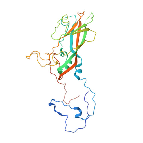
Echovirus 18 viral protein 1287Echovirus E18Mutation(s): 0 
EC: 3.4.22.29 (PDB Primary Data), 3.6.1.15 (PDB Primary Data), 3.4.22.28 (PDB Primary Data), 2.7.7.48 (PDB Primary Data)
UniProt
Find proteins for Q8V635 (Echovirus E18)
Explore Q8V635 
Go to UniProtKB:  Q8V635
Entity Groups  
Sequence Clusters30% Identity50% Identity70% Identity90% Identity95% Identity100% Identity
UniProt GroupQ8V635
Sequence Annotations
Expand
  • Reference Sequence
Find similar proteins by:  (by identity cutoff)  |  3D Structure
Entity ID: 2
MoleculeChains Sequence LengthOrganismDetailsImage
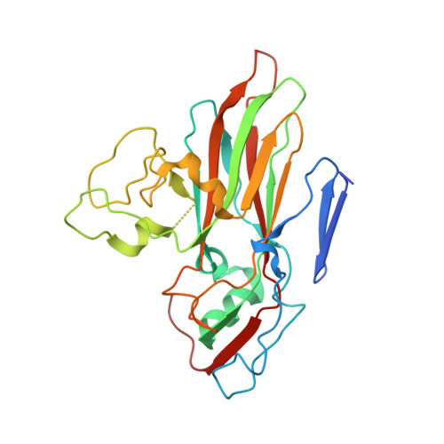
Echovirus 18 viral protein 2260Echovirus E18Mutation(s): 0 
EC: 3.4.22.29 (PDB Primary Data), 3.6.1.15 (PDB Primary Data), 3.4.22.28 (PDB Primary Data), 2.7.7.48 (PDB Primary Data)
UniProt
Find proteins for Q8V635 (Echovirus E18)
Explore Q8V635 
Go to UniProtKB:  Q8V635
Entity Groups  
Sequence Clusters30% Identity50% Identity70% Identity90% Identity95% Identity100% Identity
UniProt GroupQ8V635
Sequence Annotations
Expand
  • Reference Sequence
Find similar proteins by:  (by identity cutoff)  |  3D Structure
Entity ID: 3
MoleculeChains Sequence LengthOrganismDetailsImage
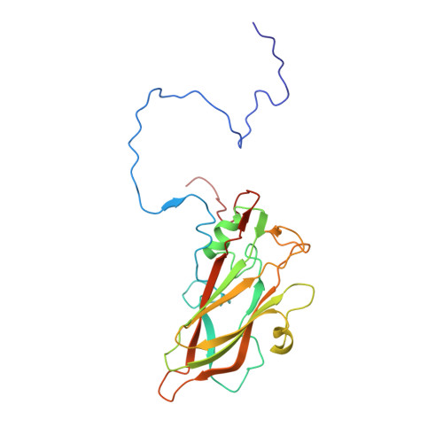
Echovirus 18 viral protein 3239Echovirus E18Mutation(s): 0 
EC: 3.4.22.29 (PDB Primary Data), 3.6.1.15 (PDB Primary Data), 3.4.22.28 (PDB Primary Data), 2.7.7.48 (PDB Primary Data)
UniProt
Find proteins for Q8V635 (Echovirus E18)
Explore Q8V635 
Go to UniProtKB:  Q8V635
Entity Groups  
Sequence Clusters30% Identity50% Identity70% Identity90% Identity95% Identity100% Identity
UniProt GroupQ8V635
Sequence Annotations
Expand
  • Reference Sequence
Find similar proteins by:  (by identity cutoff)  |  3D Structure
Entity ID: 4
MoleculeChains Sequence LengthOrganismDetailsImage
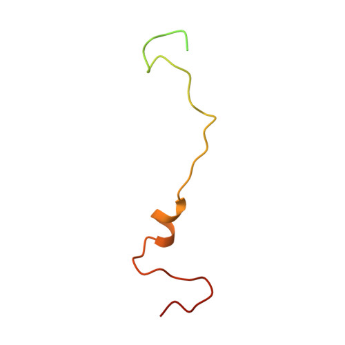
Echovirus 18 viral protein 469Echovirus E18Mutation(s): 0 
UniProt
Find proteins for Q8V635 (Echovirus E18)
Explore Q8V635 
Go to UniProtKB:  Q8V635
Entity Groups  
Sequence Clusters30% Identity50% Identity70% Identity90% Identity95% Identity100% Identity
UniProt GroupQ8V635
Sequence Annotations
Expand
  • Reference Sequence
Small Molecules
Experimental Data & Validation

Experimental Data

  • Method: ELECTRON MICROSCOPY
  • Resolution: 3.16 Å
  • Aggregation State: PARTICLE 
  • Reconstruction Method: SINGLE PARTICLE 
EM Software:
TaskSoftware PackageVersion
RECONSTRUCTIONRELION2.1
MODEL REFINEMENTPHENIX1.14

Structure Validation

View Full Validation Report


Entry History & Funding Information

Deposition Data


Funding OrganizationLocationGrant Number
European Research CouncilCzech Republic335855

Revision History  (Full details and data files)

  • Version 1.0: 2019-03-20
    Type: Initial release
  • Version 1.1: 2019-05-22
    Changes: Data collection, Derived calculations
  • Version 1.2: 2019-12-11
    Changes: Other
  • Version 1.3: 2021-08-04
    Changes: Other
  • Version 1.4: 2024-05-15
    Changes: Data collection, Database references, Derived calculations, Refinement description