6H8J | pdb_00006h8j

1.45 A resolution of Sporosarcina pasteurii urease inhibited in the presence of NBPTO


Experimental Data Snapshot

  • Method: X-RAY DIFFRACTION
  • Resolution: 1.45 Å
  • R-Value Free: 
    0.143 (Depositor), 0.143 (DCC) 
  • R-Value Work: 
    0.124 (Depositor), 0.125 (DCC) 
  • R-Value Observed: 
    0.125 (Depositor) 

Starting Model: experimental
View more details

wwPDB Validation 3D Report Full Report

Validation slider image for 6H8J

This is version 1.1 of the entry. See complete history

Literature

Insights into Urease Inhibition by N-( n-Butyl) Phosphoric Triamide through an Integrated Structural and Kinetic Approach.

Mazzei, L.Cianci, M.Contaldo, U.Ciurli, S.

(2019) J Agric Food Chem 67: 2127-2138

  • DOI: https://doi.org/10.1021/acs.jafc.8b04791
  • Primary Citation Related Structures: 
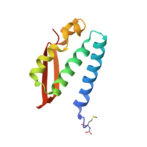
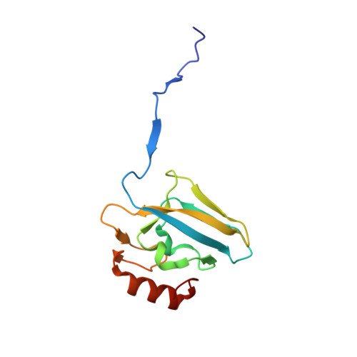
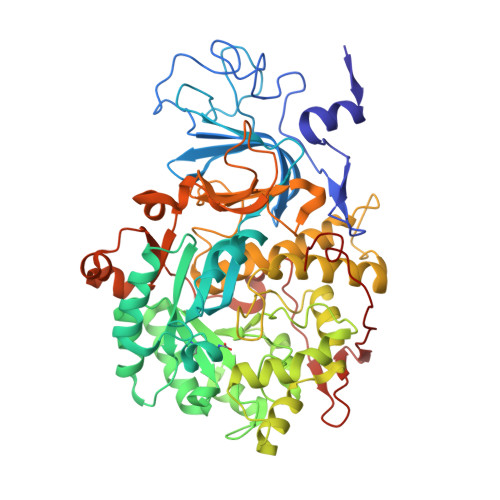
    6H8J

  • PubMed Abstract: 

    The nickel-dependent enzyme urease represents a negative element for the efficiency of soil nitrogen fertilization as well as a virulence factor for a large number of pathogenic and antibiotic-resistant bacteria. The development of ever more efficient urease inhibitors demands knowledge of their modes of action at the molecular level. N-( n-Butyl)-phosphoric triamide (NBPTO) is the oxo-derivative of N-( n-butyl)-thiophosphoric triamide (NBPT), which is extensively employed in agriculture to increase the efficiency of urea-based fertilizers. The 1.45 Å resolution structure of the enzyme-inhibitor complex obtained upon incubation of Sporosarcina pasteurii urease (SPU) with NBPTO shows the presence of diamido phosphoric acid (DAP), generated upon enzymatic hydrolysis of NBPTO with the release of n-butyl amine. DAP is bound in a tridentate binding mode to the two Ni(II) ions in the active site of urease via two O atoms and an amide NH 2 group, whereas the second amide group of DAP points away from the metal center into the active-site channel. The mobile flap modulating the size of the active-site cavity is found in a disordered closed-open conformation. A kinetic characterization of the NBPTO-based inhibition of both bacterial (SPU) and plant ( Canavalia ensiformis or jack bean, JBU) ureases, carried out by calorimetric measurements, indicates the occurrence of a reversible slow-inhibition mode of action. The latter is characterized by a very small value of the equilibrium dissociation constant of the urease-DAP complex caused, in turn, by the large rate constant for the formation of the enzyme-inhibitor complex. The much greater capability of NBPTO to inhibit urease, as compared with that of NBPT, is thus not caused by the presence of a P═O moiety versus a P═S moiety, as previously suggested, but rather by the readiness of NBPTO to react with urease without the need to convert one of the P-NH 2 amide moieties to its P-OH acid derivative, as in the case of NBPT. The latter process is indeed characterized by a very small equilibrium constant that reduces drastically the concentration of the active form of the inhibitor in the case of NBPT. This indicates that high-efficiency phosphoramide-based urease inhibitors must have at least one O atom bound to the central P atom in order for the molecule to efficiently and rapidly bind to the dinickel center of the enzyme.


  • Organizational Affiliation
    • Laboratory of Bioinorganic Chemistry, Department of Pharmacy and Biotechnology , University of Bologna , 40126 Bologna , Italy.

Macromolecule Content 

  • Total Structure Weight: 87.74 kDa 
  • Atom Count: 7,252 
  • Modeled Residue Count: 792 
  • Deposited Residue Count: 792 
  • Unique protein chains: 3

Macromolecules

Find similar proteins by:|  3D Structure
Entity ID: 1
MoleculeChains  Sequence LengthOrganismDetailsImage
Urease subunit gamma100Sporosarcina pasteuriiMutation(s): 0 
EC: 3.5.1.5
UniProt
Find proteins for P41022 (Sporosarcina pasteurii)
Explore P41022 
Go to UniProtKB:  P41022
Entity Groups
Sequence Clusters30% Identity50% Identity70% Identity90% Identity95% Identity100% Identity
UniProt GroupP41022
Sequence Annotations
Expand
Reference Sequence
Find similar proteins by:|  3D Structure
Entity ID: 2
MoleculeChains  Sequence LengthOrganismDetailsImage
Urease subunit beta122Sporosarcina pasteuriiMutation(s): 0 
EC: 3.5.1.5
UniProt
Find proteins for P41021 (Sporosarcina pasteurii)
Explore P41021 
Go to UniProtKB:  P41021
Entity Groups
Sequence Clusters30% Identity50% Identity70% Identity90% Identity95% Identity100% Identity
UniProt GroupP41021
Sequence Annotations
Expand
Reference Sequence
Find similar proteins by:|  3D Structure
Entity ID: 3
MoleculeChains  Sequence LengthOrganismDetailsImage
Urease subunit alpha570Sporosarcina pasteuriiMutation(s): 0 
EC: 3.5.1.5
UniProt
Find proteins for P41020 (Sporosarcina pasteurii)
Explore P41020 
Go to UniProtKB:  P41020
Entity Groups
Sequence Clusters30% Identity50% Identity70% Identity90% Identity95% Identity100% Identity
UniProt GroupP41020
Sequence Annotations
Expand
Reference Sequence

Small Molecules

Ligands 4 Unique
IDChains Name / Formula / InChI Key2D Diagram3D Interactions
SO4

Query on SO4


Download:Ideal Coordinates CCD File
H [auth A],
J [auth B],
K [auth B],
V [auth C],
W [auth C]
SULFATE ION
O4 S
QAOWNCQODCNURD-UHFFFAOYSA-L
2PA

Query on 2PA


Download:Ideal Coordinates CCD File
X [auth C]DIAMIDOPHOSPHATE
H5 N2 O2 P
ANCLJVISBRWUTR-UHFFFAOYSA-N
EDO

Query on EDO


Download:Ideal Coordinates CCD File
D [auth A]
E [auth A]
F [auth A]
G [auth A]
I [auth B]
D [auth A],
E [auth A],
F [auth A],
G [auth A],
I [auth B],
N [auth C],
O [auth C],
P [auth C],
Q [auth C],
R [auth C],
S [auth C],
T [auth C],
U [auth C]
1,2-ETHANEDIOL
C2 H6 O2
LYCAIKOWRPUZTN-UHFFFAOYSA-N
NI

Query on NI


Download:Ideal Coordinates CCD File
L [auth C],
M [auth C]
NICKEL (II) ION
Ni
VEQPNABPJHWNSG-UHFFFAOYSA-N
Modified Residues  2 Unique
IDChains TypeFormula2D DiagramParent
CXM
Query on CXM
A
L-PEPTIDE LINKINGC6 H11 N O4 SMET
KCX
Query on KCX
C
L-PEPTIDE LINKINGC7 H14 N2 O4LYS

Experimental Data & Validation

Experimental Data

  • Method: X-RAY DIFFRACTION
  • Resolution: 1.45 Å
  • R-Value Free:  0.143 (Depositor), 0.143 (DCC) 
  • R-Value Work:  0.124 (Depositor), 0.125 (DCC) 
  • R-Value Observed: 0.125 (Depositor) 
Space Group: P 63 2 2
Unit Cell:
Length ( Å )Angle ( ˚ )
a = 131.446α = 90
b = 131.446β = 90
c = 188.716γ = 120
Software Package:
Software NamePurpose
REFMACrefinement
XDSdata reduction
Aimlessdata scaling
REFMACphasing

Structure Validation

View Full Validation Report


Entry History 

Deposition Data

Revision History  (Full details and data files)

  • Version 1.0: 2019-06-12
    Type: Initial release
  • Version 1.1: 2024-01-17
    Changes: Data collection, Database references, Derived calculations, Refinement description