6GI5

Crystal structure of the ferric enterobactin esterase (PfeE) from Pseudomonas aeruginosa in complex with the tris-catechol vector


Experimental Data Snapshot

  • Method: X-RAY DIFFRACTION
  • Resolution: 3.11 Å
  • R-Value Free: 0.285 
  • R-Value Work: 0.242 
  • R-Value Observed: 0.244 

wwPDB Validation   3D Report Full Report


Ligand Structure Quality Assessment 


This is version 1.2 of the entry. See complete history


Literature

A Key Role for the Periplasmic PfeE Esterase in Iron Acquisition via the Siderophore Enterobactin in Pseudomonas aeruginosa.

Perraud, Q.Moynie, L.Gasser, V.Munier, M.Godet, J.Hoegy, F.Mely, Y.Mislin, G.L.A.Naismith, J.H.Schalk, I.J.

(2018) ACS Chem Biol 13: 2603-2614

  • DOI: https://doi.org/10.1021/acschembio.8b00543
  • Primary Citation of Related Structures:  
    6GI0, 6GI1, 6GI2, 6GI5

  • PubMed Abstract: 

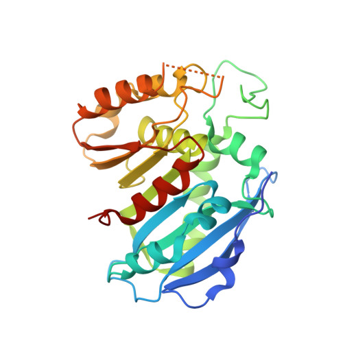
    Enterobactin (ENT) is a siderophore (iron-chelating compound) produced by Escherichia coli to gain access to iron, an indispensable nutrient for bacterial growth. ENT is used as an exosiderophore by Pseudomonas aeruginosa with transport of ferri-ENT across the outer membrane by the PfeA transporter. Next to the pfeA gene on the chromosome is localized a gene encoding for an esterase, PfeE, whose transcription is regulated, as for pfeA, by the presence of ENT in bacterial environment. Purified PfeE hydrolyzed ferri-ENT into three molecules of 2,3-DHBS (2,3-dihydroxybenzoylserine) still complexed with ferric iron, and complete dissociation of iron from ENT chelating groups was only possible in the presence of both PfeE and an iron reducer, such as DTT. The crystal structure of PfeE and an inactive PfeE mutant complexed with ferri-ENT or a nonhydrolyzable ferri-catechol complex allowed identification of the enzyme binding site and the catalytic triad. Finally, cell fractionation and fluorescence microscopy showed periplasmic localization of PfeE in P. aeruginosa cells. Thus, the molecular mechanism of iron dissociation from ENT in P. aeruginosa differs from that previously described in E. coli. In P. aeruginosa, siderophore hydrolysis occurs in the periplasm, with ENT never reaching the bacterial cytoplasm. In E. coli, ferri-ENT crosses the inner membrane via the ABC transporter FepBCD and ferri-ENT is hydrolyzed by the esterase Fes only once it is in the cytoplasm.


  • Organizational Affiliation

    Université de Strasbourg, UMR7242, ESBS , 300 Bld Sébastien Brant , F-67413 Illkirch, Strasbourg , France.


Macromolecules
Find similar proteins by:  (by identity cutoff)  |  3D Structure
Entity ID: 1
MoleculeChains Sequence LengthOrganismDetailsImage
Ferric enterobactin esterase
A, B
282Pseudomonas aeruginosaMutation(s): 0 
Gene Names: PA2689
UniProt
Find proteins for Q9I0F2 (Pseudomonas aeruginosa (strain ATCC 15692 / DSM 22644 / CIP 104116 / JCM 14847 / LMG 12228 / 1C / PRS 101 / PAO1))
Explore Q9I0F2 
Go to UniProtKB:  Q9I0F2
Entity Groups  
Sequence Clusters30% Identity50% Identity70% Identity90% Identity95% Identity100% Identity
UniProt GroupQ9I0F2
Sequence Annotations
Expand
  • Reference Sequence
Small Molecules
Ligands 2 Unique
IDChains Name / Formula / InChI Key2D Diagram3D Interactions
8SW
Query on 8SW

Download Ideal Coordinates CCD File 
D [auth A],
F [auth B]
~{N}-[2-[[(2~{S})-2-[[2,3-bis(oxidanyl)phenyl]carbonylamino]-3-[[(2~{S})-2-[[2,3-bis(oxidanyl)phenyl]carbonylamino]-3-oxidanylidene-3-(prop-2-ynylamino)propyl]amino]-3-oxidanylidene-propyl]amino]-2-oxidanylidene-ethyl]-2,3-bis(oxidanyl)benzamide
C32 H32 N6 O12
XJMZSRWAQFOMTB-PMACEKPBSA-N
FE
Query on FE

Download Ideal Coordinates CCD File 
C [auth A],
E [auth B]
FE (III) ION
Fe
VTLYFUHAOXGGBS-UHFFFAOYSA-N
Experimental Data & Validation

Experimental Data

  • Method: X-RAY DIFFRACTION
  • Resolution: 3.11 Å
  • R-Value Free: 0.285 
  • R-Value Work: 0.242 
  • R-Value Observed: 0.244 
  • Space Group: I 41 2 2
Unit Cell:
Length ( Å )Angle ( ˚ )
a = 126.612α = 90
b = 126.612β = 90
c = 293.711γ = 90
Software Package:
Software NamePurpose
REFMACrefinement
XDSdata reduction
XSCALEdata scaling
PHASERphasing

Structure Validation

View Full Validation Report


Ligand Structure Quality Assessment 


Entry History & Funding Information

Deposition Data


Funding OrganizationLocationGrant Number
United Kingdom--

Revision History  (Full details and data files)

  • Version 1.0: 2018-06-20
    Type: Initial release
  • Version 1.1: 2018-08-22
    Changes: Data collection, Database references
  • Version 1.2: 2018-10-03
    Changes: Data collection, Database references