6GEB | pdb_00006geb

X-ray structure of the Legionella pneumophila ATPase DotB


Experimental Data Snapshot

  • Method: X-RAY DIFFRACTION
  • Resolution: 3.19 Å
  • R-Value Free: 
    0.261 (Depositor), 0.260 (DCC) 
  • R-Value Work: 
    0.233 (Depositor), 0.234 (DCC) 
  • R-Value Observed: 
    0.234 (Depositor) 

Starting Models: experimental
View more details

wwPDB Validation 3D Report Full Report

Validation slider image for 6GEB

This is version 1.3 of the entry. See complete history

Literature

X-ray crystal structures of the type IVb secretion system DotB ATPases.

Prevost, M.S.Waksman, G.

(2018) Protein Sci 27: 1464-1475

  • DOI: https://doi.org/10.1002/pro.3439
  • Primary Citation Related Structures: 
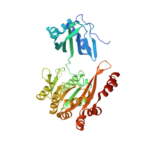
    6GEB, 6GEF

  • PubMed Abstract: 

    Human infections by the intracellular bacterial pathogen Legionella pneumophila result in a severe form of pneumonia, the Legionnaire's disease. L. pneumophila utilizes a Type IVb secretion (T4bS) system termed "dot/icm" to secrete protein effectors to the host cytoplasm. The dot/icm system is powered at least in part by a functionally critical AAA+ ATPase, a protein called DotB, thought to belong to the VirB11 family of proteins. Here we present the crystal structure of DotB at 3.19 Å resolution, in its hexameric form. We observe that DotB is in fact a structural intermediate between VirB11 and PilT family proteins, with a PAS-like N-terminal domain coupled to a RecA-like C-terminal domain. It also shares critical structural elements only found in PilT. The structure also reveals two conformers, termed α and β, with an αβαβαβ configuration. The existence of α and β conformers in this class of proteins was confirmed by solving the structure of DotB from another bacterial pathogen, Yersinia, where, intriguingly, we observed an ααβααβ configuration. The two conformers co-exist regardless of the nucleotide-bound states of the proteins. Our investigation therefore reveals that these ATPases can adopt a wider range of conformational states than was known before, shedding new light on the extraordinary spectrum of conformations these ATPases can access to carry out their function. Overall, the structure of DotB provides a template for further rational drug design to develop more specific antibiotics to tackle Legionnaire's disease. PDB Code(s): Will; be; provided.


  • Organizational Affiliation
    • Institute of Structural and Molecular Biology, University College London and Birkbeck, Malet Street, London, WC1E 7HX, United Kingdom.

Macromolecule Content 

  • Total Structure Weight: 531.09 kDa 
  • Atom Count: 34,658 
  • Modeled Residue Count: 4,420 
  • Deposited Residue Count: 4,692 
  • Unique protein chains: 1

Macromolecules

Find similar proteins by:|  3D Structure
Entity ID: 1
MoleculeChains  Sequence LengthOrganismDetailsImage
DotB
A, B, C, D, E
A, B, C, D, E, F, G, H, I, J, K, L
391Legionella pneumophilaMutation(s): 0 
Gene Names: dotByggRERS240541_01758ERS240560_02271ERS253249_00407
UniProt
Find proteins for O52185 (Legionella pneumophila)
Explore O52185 
Go to UniProtKB:  O52185
Entity Groups
Sequence Clusters30% Identity50% Identity70% Identity90% Identity95% Identity100% Identity
UniProt GroupO52185
Sequence Annotations
Expand
Reference Sequence

Small Molecules

Ligands 1 Unique
IDChains Name / Formula / InChI Key2D Diagram3D Interactions
PO4

Query on PO4


Download:Ideal Coordinates CCD File
M [auth A]
N [auth B]
O [auth C]
P [auth D]
Q [auth E]
M [auth A],
N [auth B],
O [auth C],
P [auth D],
Q [auth E],
R [auth F],
S [auth G],
T [auth H],
U [auth I],
V [auth J],
W [auth K],
X [auth L]
PHOSPHATE ION
O4 P
NBIIXXVUZAFLBC-UHFFFAOYSA-K

Experimental Data & Validation

Experimental Data

  • Method: X-RAY DIFFRACTION
  • Resolution: 3.19 Å
  • R-Value Free:  0.261 (Depositor), 0.260 (DCC) 
  • R-Value Work:  0.233 (Depositor), 0.234 (DCC) 
  • R-Value Observed: 0.234 (Depositor) 
Space Group: P 1
Unit Cell:
Length ( Å )Angle ( ˚ )
a = 109.2α = 83.7
b = 109.3β = 86.6
c = 119.8γ = 60.7
Software Package:
Software NamePurpose
REFMACrefinement
PDB_EXTRACTdata extraction
XDSdata reduction
XSCALEdata scaling
PHASERphasing

Structure Validation

View Full Validation Report


Entry History 

& Funding Information

Deposition Data


Funding OrganizationLocationGrant Number
European Research CouncilUnited Kingdom321630

Revision History  (Full details and data files)

  • Version 1.0: 2018-05-30
    Type: Initial release
  • Version 1.1: 2018-07-11
    Changes: Data collection, Database references
  • Version 1.2: 2018-10-03
    Changes: Data collection, Database references
  • Version 1.3: 2024-01-17
    Changes: Data collection, Database references, Refinement description