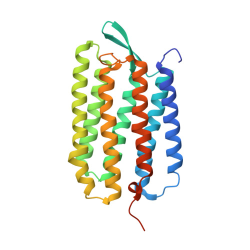
6GA8

BACTERIORHODOPSIN, 330 FS STATE, REAL-SPACE REFINED AGAINST 15% EXTRAPOLATED STRUCTURE FACTORS


Experimental Data Snapshot

  • Method: X-RAY DIFFRACTION
  • Resolution: 1.80 Å
  • R-Value Free: 0.214 
  • R-Value Work: 0.206 

wwPDB Validation   3D Report Full Report


Ligand Structure Quality Assessment 


This is version 1.1 of the entry. See complete history


Literature

Three-dimensional view of ultrafast dynamics in photoexcited bacteriorhodopsin.

Nass Kovacs, G.Colletier, J.P.Grunbein, M.L.Yang, Y.Stensitzki, T.Batyuk, A.Carbajo, S.Doak, R.B.Ehrenberg, D.Foucar, L.Gasper, R.Gorel, A.Hilpert, M.Kloos, M.Koglin, J.E.Reinstein, J.Roome, C.M.Schlesinger, R.Seaberg, M.Shoeman, R.L.Stricker, M.Boutet, S.Haacke, S.Heberle, J.Heyne, K.Domratcheva, T.Barends, T.R.M.Schlichting, I.

(2019) Nat Commun 10: 3177-3177

  • DOI: https://doi.org/10.1038/s41467-019-10758-0
  • Primary Citation of Related Structures:  
    6GA1, 6GA2, 6GA3, 6GA4, 6GA5, 6GA6, 6GA7, 6GA8, 6GA9, 6GAA, 6GAB, 6GAC, 6GAD, 6GAE, 6GAF, 6GAG, 6GAH, 6GAI, 6RMK

  • PubMed Abstract: 

    Bacteriorhodopsin (bR) is a light-driven proton pump. The primary photochemical event upon light absorption is isomerization of the retinal chromophore. Here we used time-resolved crystallography at an X-ray free-electron laser to follow the structural changes in multiphoton-excited bR from 250 femtoseconds to 10 picoseconds. Quantum chemistry and ultrafast spectroscopy were used to identify a sequential two-photon absorption process, leading to excitation of a tryptophan residue flanking the retinal chromophore, as a first manifestation of multiphoton effects. We resolve distinct stages in the structural dynamics of the all-trans retinal in photoexcited bR to a highly twisted 13-cis conformation. Other active site sub-picosecond rearrangements include correlated vibrational motions of the electronically excited retinal chromophore, the surrounding amino acids and water molecules as well as their hydrogen bonding network. These results show that this extended photo-active network forms an electronically and vibrationally coupled system in bR, and most likely in all retinal proteins.


  • Organizational Affiliation

    Max-Planck-Institut für Medizinische Forschung, Jahnstraße 29, 69120, Heidelberg, Germany.


Macromolecules
Find similar proteins by:  (by identity cutoff)  |  3D Structure
Entity ID: 1
MoleculeChains Sequence LengthOrganismDetailsImage
Bacteriorhodopsin249Halobacterium salinarum NRC-1Mutation(s): 0 
Gene Names: bopVNG_1467G
Membrane Entity: Yes 
UniProt
Find proteins for P02945 (Halobacterium salinarum (strain ATCC 700922 / JCM 11081 / NRC-1))
Explore P02945 
Go to UniProtKB:  P02945
Entity Groups  
Sequence Clusters30% Identity50% Identity70% Identity90% Identity95% Identity100% Identity
UniProt GroupP02945
Sequence Annotations
Expand
  • Reference Sequence
Small Molecules
Ligands 10 Unique
IDChains Name / Formula / InChI Key2D Diagram3D Interactions
L2P
Query on L2P

Download Ideal Coordinates CCD File 
C [auth A],
K [auth A],
L [auth A]
2,3-DI-PHYTANYL-GLYCEROL
C43 H88 O3
ISDBCJSGCHUHFI-UMZPFTBHSA-N
RET
Query on RET

Download Ideal Coordinates CCD File 
B [auth A]RETINAL
C20 H28 O
NCYCYZXNIZJOKI-OVSJKPMPSA-N
MYS
Query on MYS

Download Ideal Coordinates CCD File 
I [auth A]PENTADECANE
C15 H32
YCOZIPAWZNQLMR-UHFFFAOYSA-N
C14
Query on C14

Download Ideal Coordinates CCD File 
N [auth A]TETRADECANE
C14 H30
BGHCVCJVXZWKCC-UHFFFAOYSA-N
TRD
Query on TRD

Download Ideal Coordinates CCD File 
D [auth A]TRIDECANE
C13 H28
IIYFAKIEWZDVMP-UHFFFAOYSA-N
UND
Query on UND

Download Ideal Coordinates CCD File 
J [auth A],
P [auth A]
UNDECANE
C11 H24
RSJKGSCJYJTIGS-UHFFFAOYSA-N
D10
Query on D10

Download Ideal Coordinates CCD File 
E [auth A]DECANE
C10 H22
DIOQZVSQGTUSAI-UHFFFAOYSA-N
DD9
Query on DD9

Download Ideal Coordinates CCD File 
M [auth A]nonane
C9 H20
BKIMMITUMNQMOS-UHFFFAOYSA-N
OCT
Query on OCT

Download Ideal Coordinates CCD File 
G [auth A],
H [auth A],
O [auth A]
N-OCTANE
C8 H18
TVMXDCGIABBOFY-UHFFFAOYSA-N
HP6
Query on HP6

Download Ideal Coordinates CCD File 
F [auth A]HEPTANE
C7 H16
IMNFDUFMRHMDMM-UHFFFAOYSA-N
Experimental Data & Validation

Experimental Data

  • Method: X-RAY DIFFRACTION
  • Resolution: 1.80 Å
  • R-Value Free: 0.214 
  • R-Value Work: 0.206 
  • Space Group: P 63
Unit Cell:
Length ( Å )Angle ( ˚ )
a = 62α = 90
b = 62β = 90
c = 110.4γ = 120
Software Package:
Software NamePurpose
PHENIXrefinement
CrystFELdata reduction
CrystFELdata scaling
PHENIXphasing

Structure Validation

View Full Validation Report


Ligand Structure Quality Assessment 


Entry History & Funding Information

Revision History  (Full details and data files)

  • Version 1.0: 2019-04-24
    Type: Initial release
  • Version 1.1: 2019-07-31
    Changes: Data collection, Database references