Three-dimensional view of ultrafast dynamics in photoexcited bacteriorhodopsin.
Nass Kovacs, G., Colletier, J.P., Grunbein, M.L., Yang, Y., Stensitzki, T., Batyuk, A., Carbajo, S., Doak, R.B., Ehrenberg, D., Foucar, L., Gasper, R., Gorel, A., Hilpert, M., Kloos, M., Koglin, J.E., Reinstein, J., Roome, C.M., Schlesinger, R., Seaberg, M., Shoeman, R.L., Stricker, M., Boutet, S., Haacke, S., Heberle, J., Heyne, K., Domratcheva, T., Barends, T.R.M., Schlichting, I.(2019) Nat Commun 10: 3177-3177
- PubMed: 31320619 Search on PubMedSearch on PubMed Central
- DOI: https://doi.org/10.1038/s41467-019-10758-0
- Primary Citation Related Structures:
6GA1, 6GA2, 6GA3, 6GA4, 6GA5, 6GA6, 6GA7, 6GA8, 6GA9, 6GAA, 6GAB, 6GAC, 6GAD, 6GAE, 6GAF, 6GAG, 6GAH, 6GAI, 6RMK - PubMed Abstract:
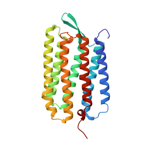
Bacteriorhodopsin (bR) is a light-driven proton pump. The primary photochemical event upon light absorption is isomerization of the retinal chromophore. Here we used time-resolved crystallography at an X-ray free-electron laser to follow the structural changes in multiphoton-excited bR from 250 femtoseconds to 10 picoseconds. Quantum chemistry and ultrafast spectroscopy were used to identify a sequential two-photon absorption process, leading to excitation of a tryptophan residue flanking the retinal chromophore, as a first manifestation of multiphoton effects. We resolve distinct stages in the structural dynamics of the all-trans retinal in photoexcited bR to a highly twisted 13-cis conformation. Other active site sub-picosecond rearrangements include correlated vibrational motions of the electronically excited retinal chromophore, the surrounding amino acids and water molecules as well as their hydrogen bonding network. These results show that this extended photo-active network forms an electronically and vibrationally coupled system in bR, and most likely in all retinal proteins.
- Max-Planck-Institut für Medizinische Forschung, Jahnstraße 29, 69120, Heidelberg, Germany.
Organizational Affiliation: