Mining the cellular inventory of pyridoxal phosphate-dependent enzymes with functionalized cofactor mimics.
Hoegl, A., Nodwell, M.B., Kirsch, V.C., Bach, N.C., Pfanzelt, M., Stahl, M., Schneider, S., Sieber, S.A.(2018) Nat Chem 10: 1234-1245
- PubMed: 30297752 Search on PubMedSearch on PubMed Central
- DOI: https://doi.org/10.1038/s41557-018-0144-2
- Primary Citation Related Structures:
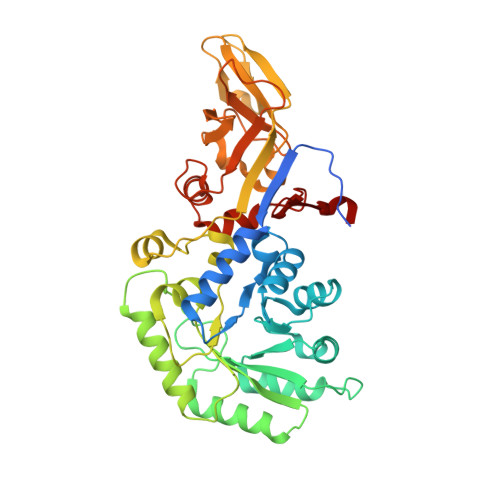
6G56, 6G58, 6G59 - PubMed Abstract:
Pyridoxal phosphate (PLP) is an enzyme cofactor required for the chemical transformation of biological amines in many central cellular processes. PLP-dependent enzymes (PLP-DEs) are ubiquitous and evolutionarily diverse, making their classification based on sequence homology challenging. Here we present a chemical proteomic method for reporting on PLP-DEs using functionalized cofactor probes. We synthesized pyridoxal analogues modified at the 2'-position, which are taken up by cells and metabolized in situ. These pyridoxal analogues are phosphorylated to functional cofactor surrogates by cellular pyridoxal kinases and bind to PLP-DEs via an aldimine bond which can be rendered irreversible by NaBH 4 reduction. Conjugation to a reporter tag enables the subsequent identification of PLP-DEs using quantitative, label-free mass spectrometry. Using these probes we accessed a significant portion of the Staphylococcus aureus PLP-DE proteome (73%) and annotate uncharacterized proteins as novel PLP-DEs. We also show that this approach can be used to study structural tolerance within PLP-DE active sites and to screen for off-targets of the PLP-DE inhibitor D-cycloserine.
- Department of Chemistry, Center for Integrated Protein Science Munich (CIPSM), Technische Universität München, Garching, Germany.
Organizational Affiliation: