6G2U | pdb_00006g2u

Crystal structure of the human glutamate dehydrogenase 2 (hGDH2)


Experimental Data Snapshot

  • Method: X-RAY DIFFRACTION
  • Resolution: 2.93 Å
  • R-Value Free: 
    0.249 (Depositor), 0.189 (DCC) 
  • R-Value Work: 
    0.193 (Depositor) 
  • R-Value Observed: 
    0.196 (Depositor) 

Starting Model: experimental
View more details

wwPDB Validation 3D Report Full Report

Validation slider image for 6G2U

This is version 1.3 of the entry. See complete history

Literature

Crystal structure of glutamate dehydrogenase 2, a positively selected novel human enzyme involved in brain biology and cancer pathophysiology.

Dimovasili, C.Fadouloglou, V.E.Kefala, A.Providaki, M.Kotsifaki, D.Kanavouras, K.Sarrou, I.Plaitakis, A.Zaganas, I.Kokkinidis, M.

(2021) J Neurochem 

  • DOI: https://doi.org/10.1111/jnc.15296
  • Primary Citation Related Structures: 
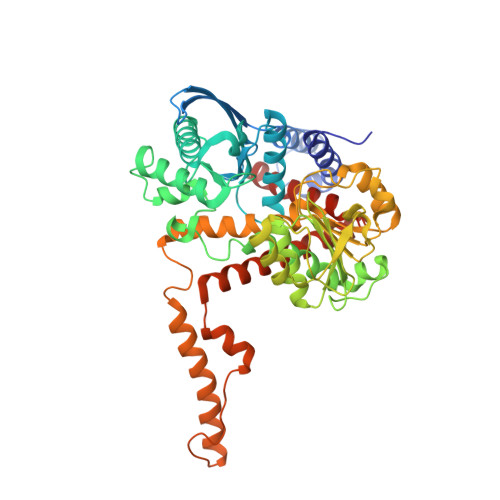
    6G2U

  • PubMed Abstract: 

    Mammalian glutamate dehydrogenase (hGDH1 in human cells) interconverts glutamate to α-ketoglutarate and ammonia while reducing NAD(P) to NAD(P)H. During primate evolution, humans and great apes have acquired hGDH2, an isoenzyme that underwent rapid evolutionary adaptation concomitantly with brain expansion, thereby acquiring unique catalytic and regulatory properties that permitted its function under conditions inhibitory to its ancestor hGDH1. Although the 3D-structures of GDHs, including hGDH1, have been determined, attempts to determine the hGDH2 structure were until recently unsuccessful. Comparison of the hGDH1/hGDH2 structures would enable a detailed understanding of their evolutionary differences. This work aimed at the determination of the hGDH2 crystal structure and the analysis of its functional implications. Recombinant hGDH2 was produced in the Spodoptera frugiperda ovarian cell line Sf21, using the Baculovirus expression system. Purification was achieved via a two-step chromatography procedure. hGDH2 was crystallized, X-ray diffraction data were collected using synchrotron radiation and the structure was determined by molecular replacement. The hGDH2 structure is reported at a resolution of 2.9 Å. The enzyme adopts a novel semi-closed conformation, which is an intermediate between known open and closed GDH1 conformations, differing from both. The structure enabled us to dissect previously reported biochemical findings and to structurally interpret the effects of evolutionary amino acid substitutions, including Arg470His, on ADP affinity. In conclusion, our data provide insights into the structural basis of hGDH2 properties, the functional evolution of hGDH isoenzymes, and open new prospects for drug design, especially for cancer therapeutics.


  • Organizational Affiliation
    • Neurology Laboratory, Medical School, University of Crete, Heraklion, Crete, Greece.

Macromolecule Content 

  • Total Structure Weight: 340.12 kDa 
  • Atom Count: 24,160 
  • Modeled Residue Count: 2,960 
  • Deposited Residue Count: 3,030 
  • Unique protein chains: 1

Macromolecules

Find similar proteins by:|  3D Structure
Entity ID: 1
MoleculeChains  Sequence LengthOrganismDetailsImage
Glutamate dehydrogenase 2, mitochondrial
A, B, C, D, E
A, B, C, D, E, F
505Homo sapiensMutation(s): 0 
Gene Names: GLUD2GLUDP1
EC: 1.4.1.3
UniProt & NIH Common Fund Data Resources
Find proteins for P49448 (Homo sapiens)
Explore P49448 
Go to UniProtKB:  P49448
PHAROS:  P49448
GTEx:  ENSG00000182890 
Entity Groups
Sequence Clusters30% Identity50% Identity70% Identity90% Identity95% Identity100% Identity
UniProt GroupP49448
Sequence Annotations
Expand
Reference Sequence

Small Molecules

Ligands 3 Unique
IDChains Name / Formula / InChI Key2D Diagram3D Interactions
PO4

Query on PO4


Download:Ideal Coordinates CCD File
AA [auth C]
FA [auth D]
G [auth A]
GA [auth D]
H [auth A]
AA [auth C],
FA [auth D],
G [auth A],
GA [auth D],
H [auth A],
I [auth A],
IB [auth F],
J [auth A],
JB [auth F],
KB [auth F],
LB [auth F],
MB [auth F],
P [auth B],
Q [auth B],
R [auth B],
S [auth B],
T [auth B],
W [auth C],
X [auth C],
XA [auth E],
Y [auth C],
YA [auth E],
Z [auth C]
PHOSPHATE ION
O4 P
NBIIXXVUZAFLBC-UHFFFAOYSA-K
CL

Query on CL


Download:Ideal Coordinates CCD File
CB [auth E]
DA [auth C]
DB [auth E]
EA [auth C]
EB [auth E]
CB [auth E],
DA [auth C],
DB [auth E],
EA [auth C],
EB [auth E],
FB [auth E],
GB [auth E],
HB [auth E],
M [auth A],
N [auth A],
O [auth A],
QB [auth F],
SA [auth D],
TA [auth D],
U [auth B],
UA [auth D],
V [auth B],
VA [auth D],
WA [auth D]
CHLORIDE ION
Cl
VEXZGXHMUGYJMC-UHFFFAOYSA-M
NA

Query on NA


Download:Ideal Coordinates CCD File
AB [auth E]
BA [auth C]
BB [auth E]
CA [auth C]
HA [auth D]
AB [auth E],
BA [auth C],
BB [auth E],
CA [auth C],
HA [auth D],
IA [auth D],
JA [auth D],
K [auth A],
KA [auth D],
L [auth A],
LA [auth D],
MA [auth D],
NA [auth D],
NB [auth F],
OA [auth D],
OB [auth F],
PA [auth D],
PB [auth F],
QA [auth D],
RA [auth D],
ZA [auth E]
SODIUM ION
Na
FKNQFGJONOIPTF-UHFFFAOYSA-N

Experimental Data & Validation

Experimental Data

  • Method: X-RAY DIFFRACTION
  • Resolution: 2.93 Å
  • R-Value Free:  0.249 (Depositor), 0.189 (DCC) 
  • R-Value Work:  0.193 (Depositor) 
  • R-Value Observed: 0.196 (Depositor) 
Space Group: C 2 2 21
Unit Cell:
Length ( Å )Angle ( ˚ )
a = 121.795α = 90
b = 149.304β = 90
c = 433.315γ = 90
Software Package:
Software NamePurpose
PHENIXrefinement
PHENIXrefinement
XDSdata reduction
XDSdata scaling
PHASERphasing

Structure Validation

View Full Validation Report


Entry History 

& Funding Information

Deposition Data


Funding OrganizationLocationGrant Number
European CommissionGreece316223

Revision History  (Full details and data files)

  • Version 1.0: 2019-04-10
    Type: Initial release
  • Version 1.1: 2019-11-06
    Changes: Data collection, Refinement description
  • Version 1.2: 2021-02-10
    Changes: Database references, Derived calculations
  • Version 1.3: 2024-01-17
    Changes: Data collection, Database references, Refinement description