6G28

Human [protein ADP-ribosylargenine] hydrolase ARH1 in complex with ADP-ribose


Experimental Data Snapshot

  • Method: X-RAY DIFFRACTION
  • Resolution: 1.23 Å
  • R-Value Free: 0.157 
  • R-Value Work: 0.145 
  • R-Value Observed: 0.146 

wwPDB Validation   3D Report Full Report


Ligand Structure Quality Assessment 


This is version 1.3 of the entry. See complete history


Literature

(ADP-ribosyl)hydrolases: Structural Basis for Differential Substrate Recognition and Inhibition.

Rack, J.G.M.Ariza, A.Drown, B.S.Henfrey, C.Bartlett, E.Shirai, T.Hergenrother, P.J.Ahel, I.

(2018) Cell Chem Biol 25: 1533-1546.e12

  • DOI: https://doi.org/10.1016/j.chembiol.2018.11.001
  • Primary Citation of Related Structures:  
    6G1P, 6G1Q, 6G28, 6G2A, 6HGZ, 6HH3, 6HH4, 6HH5, 6HH6, 6HOZ

  • PubMed Abstract: 

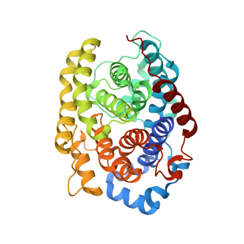
    Protein ADP-ribosylation is a highly dynamic post-translational modification. The rapid turnover is achieved, among others, by ADP-(ribosyl)hydrolases (ARHs), an ancient family of enzymes that reverses this modification. Recently ARHs came into focus due to their role as regulators of cellular stresses and tumor suppressors. Here we present a comprehensive structural analysis of the enzymatically active family members ARH1 and ARH3. These two enzymes have very distinct substrate requirements. Our data show that binding of the adenosine ribose moiety is highly diverged between the two enzymes, whereas the active sites harboring the distal ribose closely resemble each other. Despite this apparent similarity, we elucidate the structural basis for the selective inhibition of ARH3 by the ADP-ribose analogues ADP-HPD and arginine-ADP-ribose. Together, our biochemical and structural work provides important insights into the mode of enzyme-ligand interaction, helps to understand differences in their catalytic behavior, and provides useful tools for targeted drug design.


  • Organizational Affiliation

    Sir William Dunn School of Pathology, Oxford University, South Parks Road, Oxford OX1 3RE, UK.


Macromolecules
Find similar proteins by:  (by identity cutoff)  |  3D Structure
Entity ID: 1
MoleculeChains Sequence LengthOrganismDetailsImage
[Protein ADP-ribosylarginine] hydrolase367Homo sapiensMutation(s): 0 
Gene Names: ADPRHARH1
EC: 3.2.2.19
UniProt & NIH Common Fund Data Resources
Find proteins for P54922 (Homo sapiens)
Explore P54922 
Go to UniProtKB:  P54922
PHAROS:  P54922
GTEx:  ENSG00000144843 
Entity Groups  
Sequence Clusters30% Identity50% Identity70% Identity90% Identity95% Identity100% Identity
UniProt GroupP54922
Sequence Annotations
Expand
  • Reference Sequence
Experimental Data & Validation

Experimental Data

  • Method: X-RAY DIFFRACTION
  • Resolution: 1.23 Å
  • R-Value Free: 0.157 
  • R-Value Work: 0.145 
  • R-Value Observed: 0.146 
  • Space Group: P 21 21 21
Unit Cell:
Length ( Å )Angle ( ˚ )
a = 64.102α = 90
b = 66.687β = 90
c = 83.42γ = 90
Software Package:
Software NamePurpose
REFMACrefinement
xia2data reduction
DIALSdata scaling
PHASERphasing

Structure Validation

View Full Validation Report


Ligand Structure Quality Assessment 


Entry History & Funding Information

Deposition Data

  • Released Date: 2018-11-28 
  • Deposition Author(s): Ariza, A.

Funding OrganizationLocationGrant Number
Wellcome TrustUnited Kingdom101794

Revision History  (Full details and data files)

  • Version 1.0: 2018-11-28
    Type: Initial release
  • Version 1.1: 2018-12-05
    Changes: Data collection, Database references
  • Version 1.2: 2019-01-02
    Changes: Data collection, Database references
  • Version 1.3: 2024-01-17
    Changes: Data collection, Database references, Derived calculations, Refinement description, Structure summary