6FLU

Photorhabdus asymbiotica lectin (PHL) in complex with synthetic C-fucoside


Experimental Data Snapshot

  • Method: X-RAY DIFFRACTION
  • Resolution: 1.78 Å
  • R-Value Free: 0.222 
  • R-Value Work: 0.184 
  • R-Value Observed: 0.186 

wwPDB Validation   3D Report Full Report


Ligand Structure Quality Assessment 


This is version 1.1 of the entry. See complete history


Literature

Fucosylated inhibitors of recently identified bangle lectin from Photorhabdus asymbiotica.

Paulikova, G.Houser, J.Kasakova, M.Oroszova, B.Bertolotti, B.Parkan, K.Moravcova, J.Wimmerova, M.

(2019) Sci Rep 9: 14904-14904

  • DOI: https://doi.org/10.1038/s41598-019-51357-9
  • Primary Citation of Related Structures:  
    6FHX, 6FHY, 6FLU

  • PubMed Abstract: 

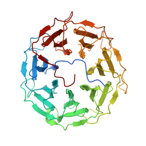
    A recently described bangle lectin (PHL) from the bacterium Photorhabdus asymbiotica was identified as a mainly fucose-binding protein that could play an important role in the host-pathogen interaction and in the modulation of host immune response. Structural studies showed that PHL is a homo-dimer that contains up to seven L-fucose-specific binding sites per monomer. For these reasons, potential ligands of the PHL lectin: α-L-fucopyranosyl-containing mono-, di-, tetra-, hexa- and dodecavalent ligands were tested. Two types of polyvalent structures were investigated - calix[4]arenes and dendrimers. The shared feature of all these structures was a C-glycosidic bond instead of the more common but physiologically unstable O-glycosidic bond. The inhibition potential of the tested structures was assessed using different techniques - hemagglutination, surface plasmon resonance, isothermal titration calorimetry, and cell cross-linking. All the ligands proved to be better than free L-fucose. The most active hexavalent dendrimer exhibited affinity three orders of magnitude higher than that of standard L-fucose. To determine the binding mode of some ligands, crystal complex PHL/fucosides 2 - 4 were prepared and studied using X-ray crystallography. The electron density in complexes proved the presence of the compounds in 6 out of 7 fucose-binding sites.


  • Organizational Affiliation

    Central European Institute of Technology, Masaryk University, Brno, Czech Republic.


Macromolecules
Find similar proteins by:  (by identity cutoff)  |  3D Structure
Entity ID: 1
MoleculeChains Sequence LengthOrganismDetailsImage
Photorhabdus asymbitoca lectin PHL
A, B
369Photorhabdus asymbiotica subsp. asymbiotica ATCC 43949Mutation(s): 0 
Gene Names: PAU_00698
UniProt
Find proteins for C7BLE4 (Photorhabdus asymbiotica subsp. asymbiotica (strain ATCC 43949 / 3105-77))
Explore C7BLE4 
Go to UniProtKB:  C7BLE4
Entity Groups  
Sequence Clusters30% Identity50% Identity70% Identity90% Identity95% Identity100% Identity
UniProt GroupC7BLE4
Sequence Annotations
Expand
  • Reference Sequence
Small Molecules
Ligands 3 Unique
IDChains Name / Formula / InChI Key2D Diagram3D Interactions
DTK
Query on DTK

Download Ideal Coordinates CCD File 
C [auth A]
D [auth A]
E [auth A]
F [auth A]
G [auth A]
C [auth A],
D [auth A],
E [auth A],
F [auth A],
G [auth A],
H [auth A],
N [auth B],
O [auth B],
P [auth B],
Q [auth B],
R [auth B],
S [auth B]
(2~{S},3~{S},4~{R},5~{S},6~{S})-2-[[(2~{R},3~{S},4~{R},6~{S})-2-(hydroxymethyl)-6-methoxy-3-oxidanyl-oxan-4-yl]methyl]-6-methyl-oxane-3,4,5-triol
C14 H26 O8
ZKQNJYZBKMAROQ-UDZHPATASA-N
CL
Query on CL

Download Ideal Coordinates CCD File 
J [auth A]
K [auth A]
L [auth A]
M [auth A]
V [auth B]
J [auth A],
K [auth A],
L [auth A],
M [auth A],
V [auth B],
W [auth B]
CHLORIDE ION
Cl
VEXZGXHMUGYJMC-UHFFFAOYSA-M
NA
Query on NA

Download Ideal Coordinates CCD File 
I [auth A],
T [auth B],
U [auth B]
SODIUM ION
Na
FKNQFGJONOIPTF-UHFFFAOYSA-N
Experimental Data & Validation

Experimental Data

  • Method: X-RAY DIFFRACTION
  • Resolution: 1.78 Å
  • R-Value Free: 0.222 
  • R-Value Work: 0.184 
  • R-Value Observed: 0.186 
  • Space Group: P 32 2 1
Unit Cell:
Length ( Å )Angle ( ˚ )
a = 81.94α = 90
b = 81.94β = 90
c = 224.645γ = 120
Software Package:
Software NamePurpose
REFMACrefinement
XDSdata reduction
SCALAdata scaling
PHASERphasing

Structure Validation

View Full Validation Report


Ligand Structure Quality Assessment 


Entry History 

Deposition Data

Revision History  (Full details and data files)

  • Version 1.0: 2019-02-06
    Type: Initial release
  • Version 1.1: 2019-10-30
    Changes: Data collection, Database references