6FDE

Crystal Structure of the HHD2 Domain of Whirlin : 3-helix conformation


Experimental Data Snapshot

  • Method: X-RAY DIFFRACTION
  • Resolution: 1.85 Å
  • R-Value Free: 0.257 
  • R-Value Work: 0.214 
  • R-Value Observed: 0.219 

wwPDB Validation   3D Report Full Report


This is version 1.1 of the entry. See complete history


Literature

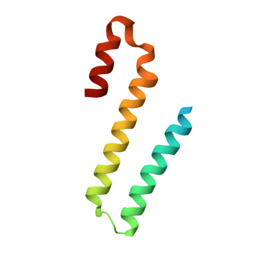
Structural plasticity of the HHD2 domain of whirlin.

Delhommel, F.Cordier, F.Saul, F.Chataigner, L.Haouz, A.Wolff, N.

(2018) FEBS J 285: 3738-3752

  • DOI: https://doi.org/10.1111/febs.14614
  • Primary Citation of Related Structures:  
    6FDD, 6FDE

  • PubMed Abstract: 

    Whirlin is a protein essential to sensory neurons. Its defects are responsible for nonsyndromic deafness or for the Usher syndrome, a condition associating congenital deafness and progressive blindness. This large multidomain scaffolding protein is expressed in three isoforms with different functions and localizations in stereocilia bundles of hearing hair cells or in the connecting cilia of photoreceptor cells. The HHD2 domain of whirlin is the only domain shared by all isoforms, but its function remains unknown. In this article, we report its crystal structure in two distinct conformations, a monomeric five-helix bundle, similar to the known structure of other HHD domains, and a three-helix bundle organized as a swapped dimer. Most of the hydrophobic contacts and electrostatic interactions that maintain the globular monomeric form are conserved at the protomer interface of the dimer. NMR experiments revealed that the five-helix conformation is predominant in solution, but exhibits increased dynamics on one face encompassing the hinge loops. Using NMR and SAXS, we also show that HHD2 does not interact with its preceding domains. Our findings suggest that structural plasticity might play a role in the function of the HHD2 domain.


  • Organizational Affiliation

    Unité Récepteurs-Canaux, Institut Pasteur, Paris, France.


Macromolecules
Find similar proteins by:  (by identity cutoff)  |  3D Structure
Entity ID: 1
MoleculeChains Sequence LengthOrganismDetailsImage
Whirlin85Mus musculusMutation(s): 0 
Gene Names: WhrnDfnb31Kiaa1526
UniProt & NIH Common Fund Data Resources
Find proteins for Q80VW5 (Mus musculus)
Explore Q80VW5 
Go to UniProtKB:  Q80VW5
IMPC:  MGI:2682003
Entity Groups  
Sequence Clusters30% Identity50% Identity70% Identity90% Identity95% Identity100% Identity
UniProt GroupQ80VW5
Sequence Annotations
Expand
  • Reference Sequence
Experimental Data & Validation

Experimental Data

  • Method: X-RAY DIFFRACTION
  • Resolution: 1.85 Å
  • R-Value Free: 0.257 
  • R-Value Work: 0.214 
  • R-Value Observed: 0.219 
  • Space Group: P 41 21 2
Unit Cell:
Length ( Å )Angle ( ˚ )
a = 34.668α = 90
b = 34.668β = 90
c = 99.732γ = 90
Software Package:
Software NamePurpose
BUSTERrefinement
XDSdata reduction
XSCALEdata scaling
PHASERphasing

Structure Validation

View Full Validation Report


Entry History & Funding Information

Deposition Data


Funding OrganizationLocationGrant Number
Fondation de France-Committee Berthe FouassierFrance--
Institut PasteurFrance--
Ministere de l'Enseignement Superieur et de la RechercheFrance--

Revision History  (Full details and data files)

  • Version 1.0: 2018-08-08
    Type: Initial release
  • Version 1.1: 2018-10-31
    Changes: Data collection, Database references