6F3H

Crystal structure of Dss1 exoribonuclease active site mutant D477N from Candida glabrata


Experimental Data Snapshot

  • Method: X-RAY DIFFRACTION
  • Resolution: 2.70 Å
  • R-Value Free: 0.240 
  • R-Value Work: 0.196 
  • R-Value Observed: 0.199 

wwPDB Validation   3D Report Full Report


This is version 1.0 of the entry. See complete history


Literature

Structural analysis of mtEXO mitochondrial RNA degradosome reveals tight coupling of nuclease and helicase components.

Razew, M.Warkocki, Z.Taube, M.Kolondra, A.Czarnocki-Cieciura, M.Nowak, E.Labedzka-Dmoch, K.Kawinska, A.Piatkowski, J.Golik, P.Kozak, M.Dziembowski, A.Nowotny, M.

(2018) Nat Commun 9: 97-97

  • DOI: https://doi.org/10.1038/s41467-017-02570-5
  • Primary Citation of Related Structures:  
    6F3H, 6F4A

  • PubMed Abstract: 

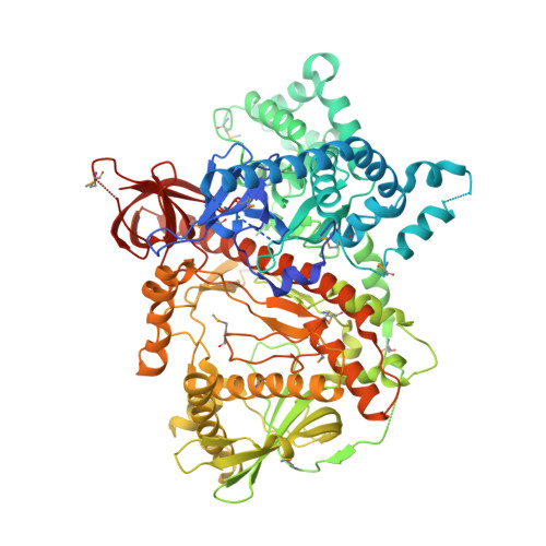
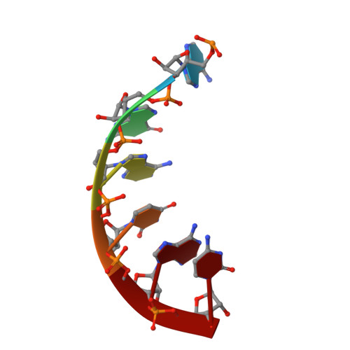
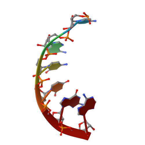
    Nuclease and helicase activities play pivotal roles in various aspects of RNA processing and degradation. These two activities are often present in multi-subunit complexes from nucleic acid metabolism. In the mitochondrial exoribonuclease complex (mtEXO) both enzymatic activities are tightly coupled making it an excellent minimal system to study helicase-exoribonuclease coordination. mtEXO is composed of Dss1 3'-to-5' exoribonuclease and Suv3 helicase. It is the master regulator of mitochondrial gene expression in yeast. Here, we present the structure of mtEXO and a description of its mechanism of action. The crystal structure of Dss1 reveals domains that are responsible for interactions with Suv3. Importantly, these interactions are compatible with the conformational changes of Suv3 domains during the helicase cycle. We demonstrate that mtEXO is an intimate complex which forms an RNA-binding channel spanning its entire structure, with Suv3 helicase feeding the 3' end of the RNA toward the active site of Dss1.


  • Organizational Affiliation

    Laboratory of Protein Structure, International Institute of Molecular and Cell Biology, Trojdena 4, 02-109, Warsaw, Poland.


Macromolecules

Find similar proteins by:  (by identity cutoff)  |  3D Structure
Entity ID: 1
MoleculeChains Sequence LengthOrganismDetailsImage
Exoribonuclease II, mitochondrial
A, B
832Nakaseomyces glabratusMutation(s): 1 
Gene Names: CAGL0M07051g
UniProt
Find proteins for Q6FJE0 (Candida glabrata (strain ATCC 2001 / BCRC 20586 / JCM 3761 / NBRC 0622 / NRRL Y-65 / CBS 138))
Explore Q6FJE0 
Go to UniProtKB:  Q6FJE0
Entity Groups  
Sequence Clusters30% Identity50% Identity70% Identity90% Identity95% Identity100% Identity
UniProt GroupQ6FJE0
Sequence Annotations
Expand
  • Reference Sequence

Find similar nucleic acids by:  Sequence   |   3D Structure  

Entity ID: 2
MoleculeChains LengthOrganismImage
RNA (5'-R(P*AP*GP*AP*UP*AP*C)-3')6Escherichia coli BL21(DE3)
Sequence Annotations
Expand
  • Reference Sequence

Find similar nucleic acids by:  Sequence   |   3D Structure  

Entity ID: 3
MoleculeChains LengthOrganismImage
RNA (5'-R(P*CP*AP*CP*UP*GP*A)-3')6Escherichia coli BL21(DE3)
Sequence Annotations
Expand
  • Reference Sequence
Small Molecules
Modified Residues  1 Unique
IDChains TypeFormula2D DiagramParent
MSE
Query on MSE
A, B
L-PEPTIDE LINKINGC5 H11 N O2 SeMET
Experimental Data & Validation

Experimental Data

  • Method: X-RAY DIFFRACTION
  • Resolution: 2.70 Å
  • R-Value Free: 0.240 
  • R-Value Work: 0.196 
  • R-Value Observed: 0.199 
  • Space Group: P 1
Unit Cell:
Length ( Å )Angle ( ˚ )
a = 73.37α = 106.21
b = 83.23β = 106.56
c = 110.37γ = 90.93
Software Package:
Software NamePurpose
PHENIXrefinement
XDSdata reduction
XSCALEdata scaling
PHASERphasing

Structure Validation

View Full Validation Report


Entry History & Funding Information

Deposition Data


Funding OrganizationLocationGrant Number
National Science Center PolandPoland00463

Revision History  (Full details and data files)

  • Version 1.0: 2018-01-17
    Type: Initial release