6F1C

C1rC1s complex


Experimental Data Snapshot

  • Method: X-RAY DIFFRACTION
  • Resolution: 4.20 Å
  • R-Value Free: 0.305 
  • R-Value Work: 0.247 
  • R-Value Observed: 0.250 

wwPDB Validation   3D Report Full Report


Ligand Structure Quality Assessment 


This is version 2.1 of the entry. See complete history


Literature

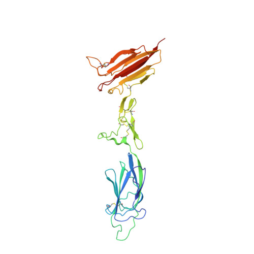
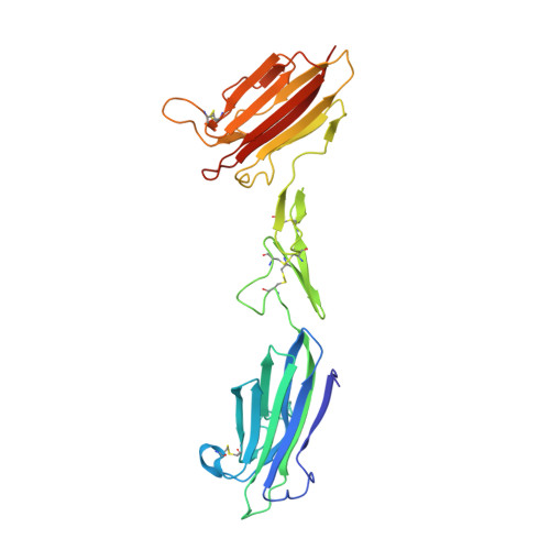
Structure of the C1r-C1s interaction of the C1 complex of complement activation.

Almitairi, J.O.M.Venkatraman Girija, U.Furze, C.M.Simpson-Gray, X.Badakshi, F.Marshall, J.E.Schwaeble, W.J.Mitchell, D.A.Moody, P.C.E.Wallis, R.

(2018) Proc Natl Acad Sci U S A 115: 768-773

  • DOI: https://doi.org/10.1073/pnas.1718709115
  • Primary Citation of Related Structures:  
    6F1C, 6F1D, 6F1H, 6F39

  • PubMed Abstract: 

    The multiprotein complex C1 initiates the classical pathway of complement activation on binding to antibody-antigen complexes, pathogen surfaces, apoptotic cells, and polyanionic structures. It is formed from the recognition subcomponent C1q and a tetramer of proteases C1r 2 C1s 2 as a Ca 2+ -dependent complex. Here we have determined the structure of a complex between the CUB1-EGF-CUB2 fragments of C1r and C1s to reveal the C1r-C1s interaction that forms the core of C1. Both fragments are L-shaped and interlock to form a compact antiparallel heterodimer with a Ca 2+ from each subcomponent at the interface. Contacts, involving all three domains of each protease, are more extensive than those of C1r or C1s homodimers, explaining why heterocomplexes form preferentially. The available structural and biophysical data support a model of C1r 2 C1s 2 in which two C1r-C1s dimers are linked via the catalytic domains of C1r. They are incompatible with a recent model in which the N-terminal domains of C1r and C1s form a fixed tetramer. On binding to C1q, the proteases become more compact, with the C1r-C1s dimers at the center and the six collagenous stems of C1q arranged around the perimeter. Activation is likely driven by separation of the C1r-C1s dimer pairs when C1q binds to a surface. Considerable flexibility in C1s likely facilitates C1 complex formation, activation of C1s by C1r, and binding and activation of downstream substrates C4 and C4b-bound C2 to initiate the reaction cascade.


  • Organizational Affiliation

    Department of Infection, Immunity, and Inflammation, University of Leicester, Leicester LE1 9HN, United Kingdom.


Macromolecules
Find similar proteins by:  (by identity cutoff)  |  3D Structure
Entity ID: 1
MoleculeChains Sequence LengthOrganismDetailsImage
Complement C1r subcomponentA,
B [auth C]
291Homo sapiensMutation(s): 0 
Gene Names: C1R
EC: 3.4.21.41
UniProt & NIH Common Fund Data Resources
Find proteins for P00736 (Homo sapiens)
Explore P00736 
Go to UniProtKB:  P00736
PHAROS:  P00736
Entity Groups  
Sequence Clusters30% Identity50% Identity70% Identity90% Identity95% Identity100% Identity
UniProt GroupP00736
Sequence Annotations
Expand
  • Reference Sequence
Find similar proteins by:  (by identity cutoff)  |  3D Structure
Entity ID: 2
MoleculeChains Sequence LengthOrganismDetailsImage
Complement C1s subcomponentC [auth D],
D [auth B]
277Homo sapiensMutation(s): 0 
Gene Names: C1S
EC: 3.4.21.42
UniProt & NIH Common Fund Data Resources
Find proteins for P09871 (Homo sapiens)
Explore P09871 
Go to UniProtKB:  P09871
PHAROS:  P09871
GTEx:  ENSG00000182326 
Entity Groups  
Sequence Clusters30% Identity50% Identity70% Identity90% Identity95% Identity100% Identity
UniProt GroupP09871
Sequence Annotations
Expand
  • Reference Sequence
Oligosaccharides

Help

Entity ID: 3
MoleculeChains Length2D Diagram Glycosylation3D Interactions
beta-D-galactopyranose-(1-4)-alpha-D-mannopyranose-(1-3)-[alpha-D-mannopyranose-(1-6)]beta-D-mannopyranose-(1-4)-2-acetamido-2-deoxy-beta-D-glucopyranose-(1-4)-2-acetamido-2-deoxy-beta-D-glucopyranose
E
6N-Glycosylation
Glycosylation Resources
GlyTouCan:  G04562GJ
GlyCosmos:  G04562GJ
GlyGen:  G04562GJ
Entity ID: 4
MoleculeChains Length2D Diagram Glycosylation3D Interactions
alpha-D-mannopyranose-(1-3)-[alpha-D-mannopyranose-(1-6)]beta-D-mannopyranose-(1-4)-2-acetamido-2-deoxy-beta-D-glucopyranose-(1-4)-2-acetamido-2-deoxy-beta-D-glucopyranose
F
5N-Glycosylation
Glycosylation Resources
GlyTouCan:  G22768VO
GlyCosmos:  G22768VO
GlyGen:  G22768VO
Entity ID: 5
MoleculeChains Length2D Diagram Glycosylation3D Interactions
beta-D-mannopyranose-(1-4)-2-acetamido-2-deoxy-beta-D-glucopyranose-(1-4)-2-acetamido-2-deoxy-beta-D-glucopyranose
G, H
3N-Glycosylation
Glycosylation Resources
GlyTouCan:  G15407YE
GlyCosmos:  G15407YE
GlyGen:  G15407YE
Small Molecules
Ligands 3 Unique
IDChains Name / Formula / InChI Key2D Diagram3D Interactions
NAG
Query on NAG

Download Ideal Coordinates CCD File 
M [auth A],
R [auth C]
2-acetamido-2-deoxy-beta-D-glucopyranose
C8 H15 N O6
OVRNDRQMDRJTHS-FMDGEEDCSA-N
CA
Query on CA

Download Ideal Coordinates CCD File 
AA [auth B]
I [auth A]
J [auth A]
K [auth A]
N [auth C]
AA [auth B],
I [auth A],
J [auth A],
K [auth A],
N [auth C],
O [auth C],
P [auth C],
S [auth D],
U [auth D],
V [auth D],
X [auth B],
Z [auth B]
CALCIUM ION
Ca
BHPQYMZQTOCNFJ-UHFFFAOYSA-N
NA
Query on NA

Download Ideal Coordinates CCD File 
BA [auth B]
L [auth A]
Q [auth C]
T [auth D]
W [auth D]
BA [auth B],
L [auth A],
Q [auth C],
T [auth D],
W [auth D],
Y [auth B]
SODIUM ION
Na
FKNQFGJONOIPTF-UHFFFAOYSA-N
Experimental Data & Validation

Experimental Data

  • Method: X-RAY DIFFRACTION
  • Resolution: 4.20 Å
  • R-Value Free: 0.305 
  • R-Value Work: 0.247 
  • R-Value Observed: 0.250 
  • Space Group: P 21 21 21
Unit Cell:
Length ( Å )Angle ( ˚ )
a = 96.779α = 90
b = 124.265β = 90
c = 195.499γ = 90
Software Package:
Software NamePurpose
EDNAdata collection
Aimlessdata scaling
PHASERphasing
PHENIXrefinement
PDB_EXTRACTdata extraction
xia2data reduction
PHASERphasing

Structure Validation

View Full Validation Report


Ligand Structure Quality Assessment 


Entry History & Funding Information

Deposition Data


Funding OrganizationLocationGrant Number
Medical Research Council (United Kingdom)United KingdomG1000191/1

Revision History  (Full details and data files)

  • Version 1.0: 2018-01-17
    Type: Initial release
  • Version 1.1: 2018-01-31
    Changes: Author supporting evidence, Database references
  • Version 2.0: 2020-07-29
    Type: Remediation
    Reason: Carbohydrate remediation
    Changes: Advisory, Atomic model, Data collection, Derived calculations, Structure summary
  • Version 2.1: 2024-01-17
    Changes: Data collection, Database references, Derived calculations, Refinement description, Structure summary