6F0C

Cytochrome P450 TxtC employs substrate conformational switching for sequential aliphatic and aromatic thaxtomin hydroxylation


Experimental Data Snapshot

  • Method: X-RAY DIFFRACTION
  • Resolution: 1.70 Å
  • R-Value Free: 0.187 
  • R-Value Work: 0.162 
  • R-Value Observed: 0.163 

wwPDB Validation   3D Report Full Report


Ligand Structure Quality Assessment 


This is version 1.2 of the entry. See complete history


Literature

Binding of Distinct Substrate Conformations Enables Hydroxylation of Remote Sites in Thaxtomin D by Cytochrome P450 TxtC.

Alkhalaf, L.M.Barry, S.M.Rea, D.Gallo, A.Griffiths, D.Lewandowski, J.R.Fulop, V.Challis, G.L.

(2019) J Am Chem Soc 141: 216-222

  • DOI: https://doi.org/10.1021/jacs.8b08864
  • Primary Citation of Related Structures:  
    6F0B, 6F0C

  • PubMed Abstract: 

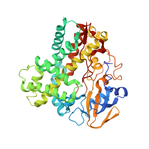
    Cytochromes P450 (CYPs) catalyze various oxidative transformations in drug metabolism, xenobiotic degradation, and natural product biosynthesis. Here we report biochemical, structural, and theoretical studies of TxtC, an unusual bifunctional CYP involved in the biosynthesis of the EPA-approved herbicide thaxtomin A. TxtC was shown to hydroxylate two remote sites within the Phe residue of its diketopiperazine substrate thaxtomin D. The reactions follow a preferred order, with hydroxylation of the α-carbon preceding functionalization of the phenyl group. To illuminate the molecular basis for remote site functionalization, X-ray crystal structures of TxtC in complex with the substrate and monohydroxylated intermediate were determined. Electron density corresponding to a diatomic molecule (probably dioxygen) was sandwiched between the heme iron atom and Thr237 in the TxtC-intermediate structure, providing insight into the mechanism for conversion of the ferrous-dioxygen complex into the reactive ferryl intermediate. The substrate and monohydroxylated intermediate adopted similar conformations in the active site, with the π-face of the phenyl group positioned over the heme iron atom. Docking simulations reproduced this observation and identified a second, energetically similar but conformationally distinct binding mode in which the α-hydrogen of the Phe residue is positioned over the heme prosthetic group. Molecular dynamics simulations confirmed that the α-hydrogen is sufficiently close to the ferryl oxygen atom to be extracted by it and indicated that the two substrate conformations cannot readily interconvert in the active site. These results indicate that TxtC is able to hydroxylate two spatially remote sites by binding distinct conformations of the substrate and monohydroxylated intermediate.


  • Organizational Affiliation

    Department of Chemistry , University of Warwick , Coventry , CV4 7AL , U.K.


Macromolecules
Find similar proteins by:  (by identity cutoff)  |  3D Structure
Entity ID: 1
MoleculeChains Sequence LengthOrganismDetailsImage
Cytochrome P450 monooxygenase395Streptomyces acidiscabies 84-104Mutation(s): 0 
Gene Names: txtCa10_01524
UniProt
Find proteins for Q8VS75 (Streptomyces acidiscabies)
Explore Q8VS75 
Go to UniProtKB:  Q8VS75
Entity Groups  
Sequence Clusters30% Identity50% Identity70% Identity90% Identity95% Identity100% Identity
UniProt GroupQ8VS75
Sequence Annotations
Expand
  • Reference Sequence
Small Molecules
Ligands 3 Unique
IDChains Name / Formula / InChI Key2D Diagram3D Interactions
HEM
Query on HEM

Download Ideal Coordinates CCD File 
B [auth A]PROTOPORPHYRIN IX CONTAINING FE
C34 H32 Fe N4 O4
KABFMIBPWCXCRK-RGGAHWMASA-L
C8B
Query on C8B

Download Ideal Coordinates CCD File 
C [auth A](3~{R},6~{S})-1,4-dimethyl-6-[(4-nitro-1~{H}-indol-3-yl)methyl]-3-oxidanyl-3-(phenylmethyl)piperazine-2,5-dione
C22 H22 N4 O5
PCCPGPMTZJOQFC-PGRDOPGGSA-N
OXY
Query on OXY

Download Ideal Coordinates CCD File 
D [auth A]OXYGEN MOLECULE
O2
MYMOFIZGZYHOMD-UHFFFAOYSA-N
Experimental Data & Validation

Experimental Data

  • Method: X-RAY DIFFRACTION
  • Resolution: 1.70 Å
  • R-Value Free: 0.187 
  • R-Value Work: 0.162 
  • R-Value Observed: 0.163 
  • Space Group: C 1 2 1
Unit Cell:
Length ( Å )Angle ( ˚ )
a = 117.4α = 90
b = 84.5β = 133.16
c = 84.4γ = 90
Software Package:
Software NamePurpose
REFMACrefinement
XDSdata reduction
XDSdata scaling
BALBESphasing

Structure Validation

View Full Validation Report


Ligand Structure Quality Assessment 


Entry History & Funding Information

Deposition Data

  • Released Date: 2018-12-12 
  • Deposition Author(s): Fulop, V.

Funding OrganizationLocationGrant Number
Biotechnology and Biological Sciences Research CouncilUnited KingdomBB/H006281/1

Revision History  (Full details and data files)

  • Version 1.0: 2018-12-12
    Type: Initial release
  • Version 1.1: 2019-07-03
    Changes: Data collection, Database references
  • Version 1.2: 2024-01-17
    Changes: Data collection, Database references, Refinement description