6EYU

Crystal structure of the inward H(+) pump xenorhodopsin


Experimental Data Snapshot

  • Method: X-RAY DIFFRACTION
  • Resolution: 2.50 Å
  • R-Value Free: 0.254 
  • R-Value Work: 0.210 
  • R-Value Observed: 0.212 

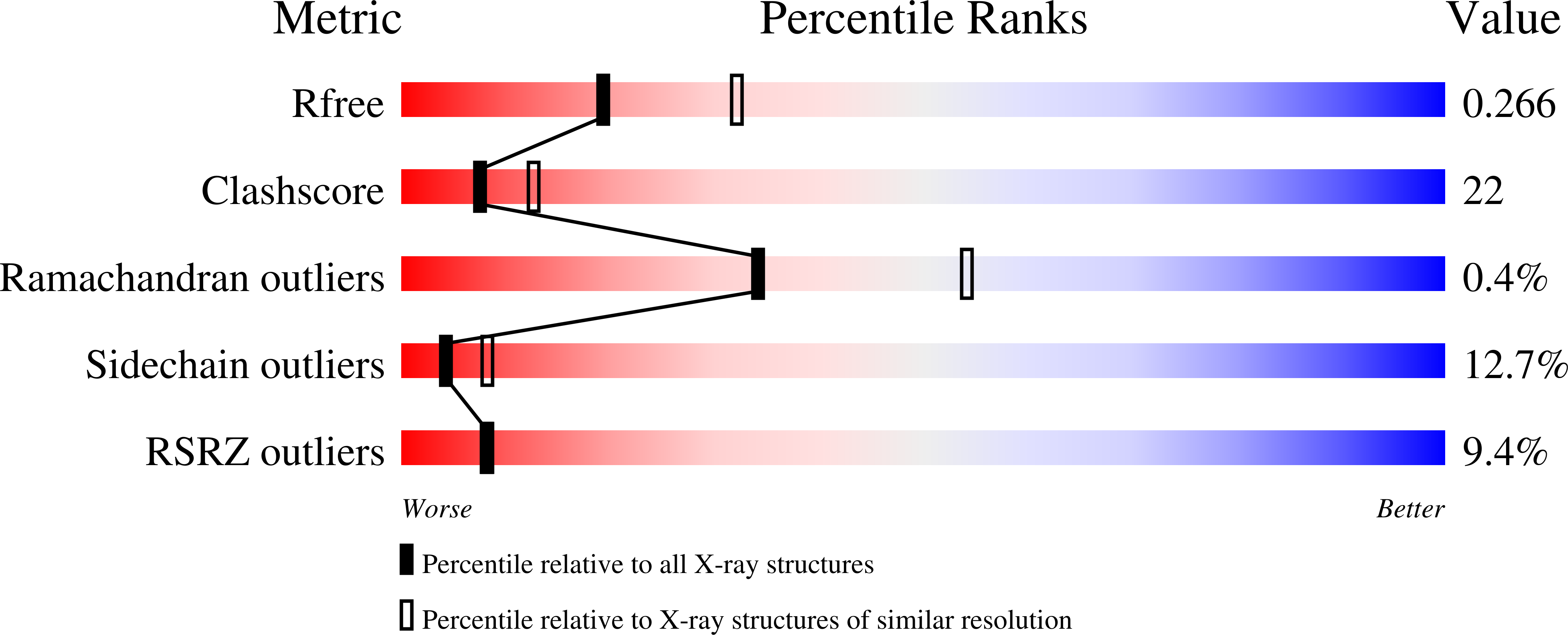
wwPDB Validation   3D Report Full Report


Ligand Structure Quality Assessment 


This is version 1.2 of the entry. See complete history


Literature

Inward H(+) pump xenorhodopsin: Mechanism and alternative optogenetic approach.

Shevchenko, V.Mager, T.Kovalev, K.Polovinkin, V.Alekseev, A.Juettner, J.Chizhov, I.Bamann, C.Vavourakis, C.Ghai, R.Gushchin, I.Borshchevskiy, V.Rogachev, A.Melnikov, I.Popov, A.Balandin, T.Rodriguez-Valera, F.Manstein, D.J.Bueldt, G.Bamberg, E.Gordeliy, V.

(2017) Sci Adv 3: e1603187

  • DOI: https://doi.org/10.1126/sciadv.1603187
  • Primary Citation of Related Structures:  
    6EYU

  • PubMed Abstract: 

    Generation of an electrochemical proton gradient is the first step of cell bioenergetics. In prokaryotes, the gradient is created by outward membrane protein proton pumps. Inward plasma membrane native proton pumps are yet unknown. We describe comprehensive functional studies of the representatives of the yet noncharacterized xenorhodopsins from Nanohaloarchaea family of microbial rhodopsins. They are inward proton pumps as we demonstrate in model membrane systems, Escherichia coli cells, human embryonic kidney cells, neuroblastoma cells, and rat hippocampal neuronal cells. We also solved the structure of a xenorhodopsin from the nanohalosarchaeon Nanosalina ( Ns XeR) and suggest a mechanism of inward proton pumping. We demonstrate that the Ns XeR is a powerful pump, which is able to elicit action potentials in rat hippocampal neuronal cells up to their maximal intrinsic firing frequency. Hence, inwardly directed proton pumps are suitable for light-induced remote control of neurons, and they are an alternative to the well-known cation-selective channelrhodopsins.


  • Organizational Affiliation

    Institute of Complex Systems (ICS), ICS-6: Structural Biochemistry, Research Centre Jülich, Jülich, Germany.


Macromolecules
Find similar proteins by:  (by identity cutoff)  |  3D Structure
Entity ID: 1
MoleculeChains Sequence LengthOrganismDetailsImage
Bacteriorhodopsin
A, B, C
232Candidatus Nanosalina sp. J07AB43Mutation(s): 0 
Gene Names: J07AB43_12860
Membrane Entity: Yes 
UniProt
Find proteins for G0QG75 (Nanosalina sp. (strain J07AB43))
Explore G0QG75 
Go to UniProtKB:  G0QG75
Entity Groups  
Sequence Clusters30% Identity50% Identity70% Identity90% Identity95% Identity100% Identity
UniProt GroupG0QG75
Sequence Annotations
Expand
  • Reference Sequence
Small Molecules
Ligands 3 Unique
IDChains Name / Formula / InChI Key2D Diagram3D Interactions
RET
Query on RET

Download Ideal Coordinates CCD File 
D [auth A],
HA [auth C],
X [auth B]
RETINAL
C20 H28 O
NCYCYZXNIZJOKI-OVSJKPMPSA-N
LFA
Query on LFA

Download Ideal Coordinates CCD File 
E [auth A]
F [auth A]
G [auth A]
H [auth A]
I [auth A]
E [auth A],
F [auth A],
G [auth A],
H [auth A],
I [auth A],
IA [auth C],
J [auth A],
JA [auth C],
KA [auth C],
LA [auth C],
MA [auth C],
Y [auth B]
EICOSANE
C20 H42
CBFCDTFDPHXCNY-UHFFFAOYSA-N
MUN
Query on MUN

Download Ideal Coordinates CCD File 
AA [auth B]
BA [auth B]
CA [auth B]
DA [auth B]
EA [auth B]
AA [auth B],
BA [auth B],
CA [auth B],
DA [auth B],
EA [auth B],
FA [auth B],
GA [auth B],
K [auth A],
L [auth A],
M [auth A],
N [auth A],
NA [auth C],
O [auth A],
OA [auth C],
P [auth A],
PA [auth C],
Q [auth A],
QA [auth C],
R [auth A],
RA [auth C],
S [auth A],
T [auth A],
U [auth A],
V [auth A],
W [auth A],
Z [auth B]
[(2~{S})-2,3-bis(oxidanyl)propyl] (~{E})-undec-2-enoate
C14 H26 O4
INMIULFREHIAOX-LXKVQUBZSA-N
Experimental Data & Validation

Experimental Data

  • Method: X-RAY DIFFRACTION
  • Resolution: 2.50 Å
  • R-Value Free: 0.254 
  • R-Value Work: 0.210 
  • R-Value Observed: 0.212 
  • Space Group: P 21 21 21
Unit Cell:
Length ( Å )Angle ( ˚ )
a = 64.01α = 90
b = 93.87β = 90
c = 196.21γ = 90
Software Package:
Software NamePurpose
XSCALEdata scaling
REFMACrefinement
PDB_EXTRACTdata extraction
XDSdata reduction
MOLREPphasing

Structure Validation

View Full Validation Report


Ligand Structure Quality Assessment 


Entry History & Funding Information

Deposition Data


Funding OrganizationLocationGrant Number
French National Research AgencyFranceANR-15-CE11-0029-02
German Research FoundationGermanyANR-15-CE11-0029-02
French Infrastructure for Integrated Structural BiologyFranceANR-10-INSB-05-02
GRAFranceANR-10-LABX-49-01
ERA.Net RUS PLUS and Ministry of Education and Science of the Russian FederationRussian FederationID 323, RFMEFI58715X0011

Revision History  (Full details and data files)

  • Version 1.0: 2017-12-13
    Type: Initial release
  • Version 1.1: 2018-01-31
    Changes: Advisory, Author supporting evidence, Derived calculations, Structure summary
  • Version 1.2: 2024-01-17
    Changes: Data collection, Database references, Refinement description