6EX7

Crystal structure of NDM-1 metallo-beta-lactamase in complex with Cd ions and a hydrolyzed beta-lactam ligand - new refinement


Experimental Data Snapshot

  • Method: X-RAY DIFFRACTION
  • Resolution: 1.95 Å
  • R-Value Free: 0.226 
  • R-Value Work: 0.195 
  • R-Value Observed: 0.196 

wwPDB Validation   3D Report Full Report


Ligand Structure Quality Assessment 


This is version 1.2 of the entry. See complete history

Re-refinement Note

This entry reflects an alternative modeling of the original data in: 4hky


Literature

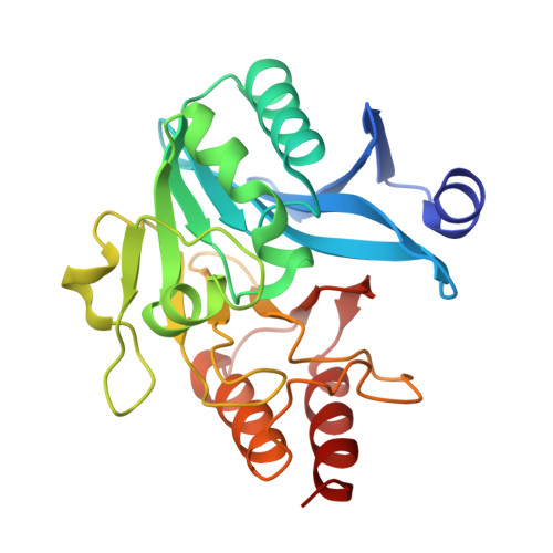
A close look onto structural models and primary ligands of metallo-beta-lactamases.

Raczynska, J.E.Shabalin, I.G.Minor, W.Wlodawer, A.Jaskolski, M.

(2018) Drug Resist Updat 40: 1-12

  • DOI: https://doi.org/10.1016/j.drup.2018.08.001
  • Primary Citation of Related Structures:  
    5N0H, 5N0I, 5NBK, 5O2E, 5O2F, 5W8W, 6EX7

  • PubMed Abstract: 

    β-Lactamases are hydrolytic enzymes capable of opening the β-lactam ring of antibiotics such as penicillin, thus endowing the bacteria that produce them with antibiotic resistance. Of particular medical concern are metallo-β-lactamases (MBLs), with an active site built around coordinated Zn cations. MBLs are pan-reactive enzymes that can break down almost all classes of β-lactams, including such last-resort antibiotics as carbapenems. They are not only broad-spectrum-reactive but are often plasmid-borne (e.g., the New Delhi enzyme, NDM), and can spread horizontally even among unrelated bacteria. Acquired MBLs are encoded by mobile genetic elements, which often include other resistance genes, making the microbiological situation particularly alarming. There is an urgent need to develop MBL inhibitors in order to rescue our antibiotic armory. A number of such efforts have been undertaken, most notably using the 3D structures of various MBLs as drug-design targets. Structure-guided drug discovery depends on the quality of the structures that are collected in the Protein Data Bank (PDB) and on the consistency of the information in dedicated β-lactamase databases. We conducted a careful review of the crystal structures of class B β-lactamases, concluding that the quality of these structures varies widely, especially in the regions where small molecules interact with the macromolecules. In a number of examples the interpretation of the bound ligands (e.g., inhibitors, substrate/product analogs) is doubtful or even incorrect, and it appears that in some cases the modeling of ligands was not supported by electron density. For ten MBL structures, alternative interpretations of the original diffraction data could be proposed and the new models have been deposited in the PDB. In four cases, these models, prepared jointly with the authors of the original depositions, superseded the previous deposits. This review emphasizes the importance of critical assessment of structural models describing key drug design targets at the level of the raw experimental data. Since the structures reviewed here are the basis for ongoing design of new MBL inhibitors, it is important to identify and correct the problems with ambiguous crystallographic interpretations, thus enhancing reproducibility in this highly medically relevant area.


  • Organizational Affiliation

    Center for Biocrystallographic Research, Institute of Bioorganic Chemistry, Polish Academy of Sciences, Poznan, Poland.


Macromolecules
Find similar proteins by:  (by identity cutoff)  |  3D Structure
Entity ID: 1
MoleculeChains Sequence LengthOrganismDetailsImage
Metallo-beta-lactamase type 2
A, B
243Klebsiella pneumoniaeMutation(s): 0 
Gene Names: blaNDM-1
EC: 3.5.2.6
UniProt
Find proteins for C7C422 (Klebsiella pneumoniae)
Explore C7C422 
Go to UniProtKB:  C7C422
Entity Groups  
Sequence Clusters30% Identity50% Identity70% Identity90% Identity95% Identity100% Identity
UniProt GroupC7C422
Sequence Annotations
Expand
  • Reference Sequence
Small Molecules
Ligands 6 Unique
IDChains Name / Formula / InChI Key2D Diagram3D Interactions
PG4
Query on PG4

Download Ideal Coordinates CCD File 
H [auth A]TETRAETHYLENE GLYCOL
C8 H18 O5
UWHCKJMYHZGTIT-UHFFFAOYSA-N
TOE
Query on TOE

Download Ideal Coordinates CCD File 
O [auth B]2-[2-(2-METHOXY-ETHOXY)-ETHOXY]-ETHOXYL
C7 H16 O4
JLGLQAWTXXGVEM-UHFFFAOYSA-N
CD
Query on CD

Download Ideal Coordinates CCD File 
C [auth A]
D [auth A]
E [auth A]
I [auth A]
J [auth B]
C [auth A],
D [auth A],
E [auth A],
I [auth A],
J [auth B],
K [auth B]
CADMIUM ION
Cd
WLZRMCYVCSSEQC-UHFFFAOYSA-N
EDO
Query on EDO

Download Ideal Coordinates CCD File 
P [auth B]1,2-ETHANEDIOL
C2 H6 O2
LYCAIKOWRPUZTN-UHFFFAOYSA-N
CL
Query on CL

Download Ideal Coordinates CCD File 
F [auth A],
L [auth B],
M [auth B]
CHLORIDE ION
Cl
VEXZGXHMUGYJMC-UHFFFAOYSA-M
UNL
Query on UNL

Download Ideal Coordinates CCD File 
G [auth A],
N [auth B]
Unknown ligand
VEXZGXHMUGYJMC-UHFFFAOYSA-M
Experimental Data & Validation

Experimental Data

  • Method: X-RAY DIFFRACTION
  • Resolution: 1.95 Å
  • R-Value Free: 0.226 
  • R-Value Work: 0.195 
  • R-Value Observed: 0.196 
  • Space Group: C 1 2 1
Unit Cell:
Length ( Å )Angle ( ˚ )
a = 145.062α = 90
b = 39.22β = 99.99
c = 75.233γ = 90
Software Package:
Software NamePurpose
REFMACrefinement
HKL-3000data reduction
HKL-3000data scaling
HKL-3000phasing
MOLREPphasing

Structure Validation

View Full Validation Report


Ligand Structure Quality Assessment 


Entry History & Funding Information

Deposition Data


Funding OrganizationLocationGrant Number
National Center for Research and DevelopmentPolandDesInMBL

Revision History  (Full details and data files)

  • Version 1.0: 2017-12-13
    Type: Initial release
  • Version 1.1: 2018-04-25
    Changes: Data collection, Database references
  • Version 1.2: 2018-12-26
    Changes: Data collection, Database references