6ETS | pdb_00006ets

Crystal structure of KDM4D with tetrazolhydrazide compound 1


Experimental Data Snapshot

  • Method: X-RAY DIFFRACTION
  • Resolution: 1.33 Å
  • R-Value Free: 
    0.156 (Depositor), 0.160 (DCC) 
  • R-Value Work: 
    0.129 (Depositor), 0.131 (DCC) 
  • R-Value Observed: 
    0.129 (Depositor) 

Starting Model: experimental
View more details

wwPDB Validation 3D Report Full Report

Validation slider image for 6ETS

This is version 1.2 of the entry. See complete history

Literature

Structure-Based Screening of Tetrazolylhydrazide Inhibitors versus KDM4 Histone Demethylases.

Malecki, P.H.Ruger, N.Roatsch, M.Krylova, O.Link, A.Jung, M.Heinemann, U.Weiss, M.S.

(2019) ChemMedChem 14: 1828-1839

  • DOI: https://doi.org/10.1002/cmdc.201900441
  • Primary Citation Related Structures: 
    6ETG, 6ETS, 6ETT, 6ETU, 6ETV, 6ETW

  • PubMed Abstract: 

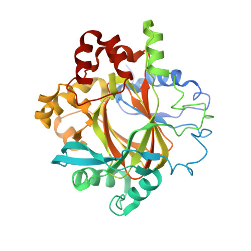
    Human histone demethylases are known to play an important role in the development of several tumor types. Consequently, they have emerged as important medical targets for the treatment of human cancer. Herein, structural studies on tetrazolylhydrazide inhibitors as a new scaffold for a certain class of histone demethylases, the JmjC proteins, are reported. A series of compounds are structurally described and their respective binding modes to the KDM4D protein, which serves as a high-resolution model to represent the KDM4 subfamily in crystallographic studies, are examined. Similar to previously reported inhibitors, the compounds described herein are competitors for the natural KDM4 cofactor, 2-oxoglutarate. The tetrazolylhydrazide scaffold fills an important gap in KDM4 inhibition and newly described, detailed interactions of inhibitor moieties pave the way to the development of compounds with high target-binding affinity and increased membrane permeability, at the same time.


  • Organizational Affiliation
    • Macromolecular Crystallography, Helmholtz-Zentrum Berlin für Materialien und Energie, Albert-Einstein-Str. 15, 12489, Berlin, Germany.

Macromolecule Content 

  • Total Structure Weight: 40.51 kDa 
  • Atom Count: 3,498 
  • Modeled Residue Count: 330 
  • Deposited Residue Count: 341 
  • Unique protein chains: 1

Macromolecules

Find similar proteins by:|  3D Structure
Entity ID: 1
MoleculeChains  Sequence LengthOrganismDetailsImage
Lysine-specific demethylase 4D341Homo sapiensMutation(s): 0 
Gene Names: KDM4DJHDM3DJMJD2D
EC: 1.14.11 (PDB Primary Data), 1.14.11.66 (UniProt)
UniProt & NIH Common Fund Data Resources
Find proteins for Q6B0I6 (Homo sapiens)
Explore Q6B0I6 
Go to UniProtKB:  Q6B0I6
PHAROS:  Q6B0I6
GTEx:  ENSG00000186280 
Entity Groups
Sequence Clusters30% Identity50% Identity70% Identity90% Identity95% Identity100% Identity
UniProt GroupQ6B0I6
Sequence Annotations
Expand
Reference Sequence

Small Molecules

Ligands 7 Unique
IDChains Name / Formula / InChI Key2D Diagram3D Interactions
I1L

Query on I1L


Download:Ideal Coordinates CCD File
Q [auth A][2-(2~{H}-1,2,3,4-tetrazol-5-yl)ethanoylamino]azanium
C3 H7 N6 O
TWKUAKLZOIABHJ-UHFFFAOYSA-O
SO4

Query on SO4


Download:Ideal Coordinates CCD File
N [auth A],
O [auth A],
P [auth A]
SULFATE ION
O4 S
QAOWNCQODCNURD-UHFFFAOYSA-L
ZN

Query on ZN


Download:Ideal Coordinates CCD File
B [auth A]ZINC ION
Zn
PTFCDOFLOPIGGS-UHFFFAOYSA-N
EDO

Query on EDO


Download:Ideal Coordinates CCD File
E [auth A]
F [auth A]
G [auth A]
H [auth A]
I [auth A]
E [auth A],
F [auth A],
G [auth A],
H [auth A],
I [auth A],
J [auth A],
K [auth A],
L [auth A],
M [auth A]
1,2-ETHANEDIOL
C2 H6 O2
LYCAIKOWRPUZTN-UHFFFAOYSA-N
NI

Query on NI


Download:Ideal Coordinates CCD File
C [auth A]NICKEL (II) ION
Ni
VEQPNABPJHWNSG-UHFFFAOYSA-N
CL

Query on CL


Download:Ideal Coordinates CCD File
R [auth A]CHLORIDE ION
Cl
VEXZGXHMUGYJMC-UHFFFAOYSA-M
NA

Query on NA


Download:Ideal Coordinates CCD File
D [auth A]SODIUM ION
Na
FKNQFGJONOIPTF-UHFFFAOYSA-N

Experimental Data & Validation

Experimental Data

  • Method: X-RAY DIFFRACTION
  • Resolution: 1.33 Å
  • R-Value Free:  0.156 (Depositor), 0.160 (DCC) 
  • R-Value Work:  0.129 (Depositor), 0.131 (DCC) 
  • R-Value Observed: 0.129 (Depositor) 
Space Group: P 43 21 2
Unit Cell:
Length ( Å )Angle ( ˚ )
a = 71.631α = 90
b = 71.631β = 90
c = 150.77γ = 90
Software Package:
Software NamePurpose
PHENIXrefinement
PDB_EXTRACTdata extraction
XDSdata reduction
XDSdata scaling
REFMACphasing

Structure Validation

View Full Validation Report


Entry History 

Deposition Data

Revision History  (Full details and data files)

  • Version 1.0: 2019-02-20
    Type: Initial release
  • Version 1.1: 2020-03-11
    Changes: Database references
  • Version 1.2: 2024-01-17
    Changes: Data collection, Database references, Derived calculations, Refinement description