6ERD | pdb_00006erd

Crystal structure of a putative acetyltransferase from Bacillus cereus species.


Experimental Data Snapshot

  • Method: X-RAY DIFFRACTION
  • Resolution: 2.00 Å
  • R-Value Free: 
    0.201 (Depositor), 0.201 (DCC) 
  • R-Value Work: 
    0.164 (Depositor), 0.164 (DCC) 
  • R-Value Observed: 
    0.166 (Depositor) 

wwPDB Validation 3D Report Full Report

Validation slider image for 6ERD

This is version 1.2 of the entry. See complete history

Literature

Functional and structural characterisation of RimL from Bacillus cereus, a new N alpha-acetyltransferase of ribosomal proteins that was wrongly assigned as an aminoglycosyltransferase.

Leonardo Silvestre, H.Asensio, J.L.Blundell, T.L.Bastida, A.Bolanos-Garcia, V.M.

(2024) Int J Biol Macromol 263: 130348-130348

  • DOI: https://doi.org/10.1016/j.ijbiomac.2024.130348
  • Primary Citation Related Structures: 
    6ERD, 8Q6U

  • PubMed Abstract: 

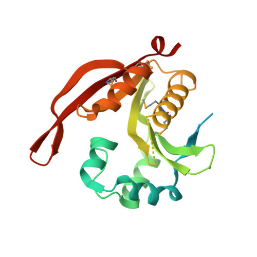
    Enzymes of the GNAT (GCN5-relate N-acetyltransferases) superfamily are important regulators of cell growth and development. They are functionally diverse and share low amino acid sequence identity, making functional annotation difficult. In this study, we report the function and structure of a new ribosomal enzyme, N α -acetyl transferase from Bacillus cereus (RimL BC ), a protein that was previously wrongly annotated as an aminoglycosyltransferase. Firstly, extensive comparative amino acid sequence analyses suggested RimL BC belongs to a cluster of proteins mediating acetylation of the ribosomal protein L7/L12. To assess if this was the case, several well established substrates of aminoglycosyltransferases were screened. The results of these studies did not support an aminoglycoside acetylating function for RimL BC . To gain further insight into RimL BC biological role, a series of studies that included MALDI-TOF, isothermal titration calorimetry, NMR, X-ray protein crystallography, and site-directed mutagenesis confirmed RimL BC affinity for Acetyl-CoA and that the ribosomal protein L7/L12 is a substrate of RimL BC . Last, we advance a mechanistic model of RimL BC mode of recognition of its protein substrates. Taken together, our studies confirmed RimL BC as a new ribosomal N α -acetyltransferase and provide structural and functional insights into substrate recognition by N α -acetyltransferases and protein acetylation in bacteria.


  • Organizational Affiliation
    • Department of Biochemistry, University of Cambridge, 80 Tennis Court Road, Cambridge CB2 1GA, United Kingdom.

Macromolecule Content 

  • Total Structure Weight: 99 kDa 
  • Atom Count: 6,149 
  • Modeled Residue Count: 662 
  • Deposited Residue Count: 844 
  • Unique protein chains: 2

Macromolecules

Find similar proteins by:|  3D Structure
Entity ID: 1
MoleculeChains  Sequence LengthOrganismDetailsImage
Aminoglycoside N6'-acetyltransferase
A, B
211Bacillus cereus ATCC 14579Mutation(s): 0 
Gene Names: BC_2494
EC: 2.3.1.82
UniProt
Find proteins for Q81D84 (Bacillus cereus (strain ATCC 14579 / DSM 31 / CCUG 7414 / JCM 2152 / NBRC 15305 / NCIMB 9373 / NCTC 2599 / NRRL B-3711))
Explore Q81D84 
Go to UniProtKB:  Q81D84
Entity Groups
Sequence Clusters30% Identity50% Identity70% Identity90% Identity95% Identity100% Identity
UniProt GroupQ81D84
Sequence Annotations
Expand
Reference Sequence
Find similar proteins by:|  3D Structure
Entity ID: 2
MoleculeChains  Sequence LengthOrganismDetailsImage
Aminoglycoside N6'-acetyltransferase
C, D
211Bacillus cereus ATCC 14579Mutation(s): 0 
Gene Names: BC_2494
EC: 2.3.1.82
UniProt
Find proteins for Q81D84 (Bacillus cereus (strain ATCC 14579 / DSM 31 / CCUG 7414 / JCM 2152 / NBRC 15305 / NCIMB 9373 / NCTC 2599 / NRRL B-3711))
Explore Q81D84 
Go to UniProtKB:  Q81D84
Entity Groups
Sequence Clusters30% Identity50% Identity70% Identity90% Identity95% Identity100% Identity
UniProt GroupQ81D84
Sequence Annotations
Expand
Reference Sequence

Small Molecules

Ligands 2 Unique
IDChains Name / Formula / InChI Key2D Diagram3D Interactions
GOL

Query on GOL


Download:Ideal Coordinates CCD File
E [auth A]
F [auth A]
G [auth A]
I [auth B]
J [auth B]
E [auth A],
F [auth A],
G [auth A],
I [auth B],
J [auth B],
K [auth B],
N [auth C],
O [auth D]
GLYCEROL
C3 H8 O3
PEDCQBHIVMGVHV-UHFFFAOYSA-N
CL

Query on CL


Download:Ideal Coordinates CCD File
H [auth A],
L [auth B],
M [auth B]
CHLORIDE ION
Cl
VEXZGXHMUGYJMC-UHFFFAOYSA-M
Modified Residues  2 Unique
IDChains TypeFormula2D DiagramParent
CME
Query on CME
A, B
L-PEPTIDE LINKINGC5 H11 N O3 S2CYS
MSE
Query on MSE
A, B
L-PEPTIDE LINKINGC5 H11 N O2 SeMET

Experimental Data & Validation

Experimental Data

  • Method: X-RAY DIFFRACTION
  • Resolution: 2.00 Å
  • R-Value Free:  0.201 (Depositor), 0.201 (DCC) 
  • R-Value Work:  0.164 (Depositor), 0.164 (DCC) 
  • R-Value Observed: 0.166 (Depositor) 
Space Group: P 1 21 1
Unit Cell:
Length ( Å )Angle ( ˚ )
a = 68.52α = 90
b = 84.17β = 98.49
c = 72.08γ = 90
Software Package:
Software NamePurpose
REFMACrefinement
xia2data reduction
SCALAdata scaling
PHENIXphasing

Structure Validation

View Full Validation Report


Entry History 

Deposition Data

Revision History  (Full details and data files)

  • Version 1.0: 2018-09-26
    Type: Initial release
  • Version 1.1: 2024-03-06
    Changes: Data collection, Database references, Refinement description
  • Version 1.2: 2024-11-13
    Changes: Structure summary