Comparison of the Binding of Reversible Inhibitors to Human Butyrylcholinesterase and Acetylcholinesterase: A Crystallographic, Kinetic and Calorimetric Study.
Rosenberry, T.L., Brazzolotto, X., Macdonald, I.R., Wandhammer, M., Trovaslet-Leroy, M., Darvesh, S., Nachon, F.(2017) Molecules 22
- PubMed: 29186056 Search on PubMedSearch on PubMed Central
- DOI: https://doi.org/10.3390/molecules22122098
- Primary Citation Related Structures:
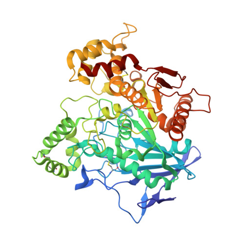
6EP4, 6EQP, 6EQQ, 6ESJ, 6ESY - PubMed Abstract:
Acetylcholinesterase (AChE) and butyrylcholinesterase (BChE) hydrolyze the neurotransmitter acetylcholine and, thereby, function as coregulators of cholinergic neurotransmission. Although closely related, these enzymes display very different substrate specificities that only partially overlap. This disparity is largely due to differences in the number of aromatic residues lining the active site gorge, which leads to large differences in the shape of the gorge and potentially to distinct interactions with an individual ligand. Considerable structural information is available for the binding of a wide diversity of ligands to AChE. In contrast, structural data on the binding of reversible ligands to BChE are lacking. In a recent effort, an inhibitor competition approach was used to probe the overlap of ligand binding sites in BChE. Here, we extend this study by solving the crystal structures of human BChE in complex with five reversible ligands, namely, decamethonium, thioflavin T, propidium, huprine, and ethopropazine. We compare these structures to equivalent AChE complexes when available in the protein data bank and supplement this comparison with kinetic data and observations from isothermal titration calorimetry. This new information now allows us to define the binding mode of various ligand families and will be of importance in designing specific reversible ligands of BChE that behave as inhibitors or reactivators.
- Departments of Neuroscience and Pharmacology, Mayo Clinic College of Medicine, Jacksonville, FL 32224, USA. rosenberry@mayo.edu.
Organizational Affiliation: