6EET

Crystal structure of mouse Protocadherin-15 EC9-MAD12

  • Classification: CELL ADHESION
  • Organism(s): Mus musculus
  • Expression System: Homo sapiens
  • Mutation(s): No 

  • Deposited: 2018-08-15 Released: 2019-08-21 
  • Deposition Author(s): Narui, Y., Sotomayor, M.
  • Funding Organization(s): National Institutes of Health/National Institute on Deafness and Other Communication Disorders (NIH/NIDCD)

Experimental Data Snapshot

  • Method: X-RAY DIFFRACTION
  • Resolution: 3.23 Å
  • R-Value Free: 0.234 
  • R-Value Work: 0.176 
  • R-Value Observed: 0.179 

wwPDB Validation   3D Report Full Report


Ligand Structure Quality Assessment 


This is version 2.2 of the entry. See complete history


Literature

Structural determinants of protocadherin-15 mechanics and function in hearing and balance perception.

Choudhary, D.Narui, Y.Neel, B.L.Wimalasena, L.N.Klanseck, C.F.De-la-Torre, P.Chen, C.Araya-Secchi, R.Tamilselvan, E.Sotomayor, M.

(2020) Proc Natl Acad Sci U S A 

  • DOI: https://doi.org/10.1073/pnas.1920444117
  • Primary Citation of Related Structures:  
    5TPK, 5ULY, 5W1D, 6BWN, 6BXU, 6E8F, 6EB5, 6EET, 6MFO, 6N22, 6N2E

  • PubMed Abstract: 

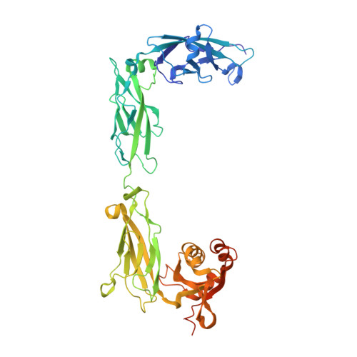
    The vertebrate inner ear, responsible for hearing and balance, is able to sense minute mechanical stimuli originating from an extraordinarily broad range of sound frequencies and intensities or from head movements. Integral to these processes is the tip-link protein complex, which conveys force to open the inner-ear transduction channels that mediate sensory perception. Protocadherin-15 and cadherin-23, two atypically large cadherins with 11 and 27 extracellular cadherin (EC) repeats, are involved in deafness and balance disorders and assemble as parallel homodimers that interact to form the tip link. Here we report the X-ray crystal structure of a protocadherin-15 + cadherin-23 heterotetrameric complex at 2.9-Å resolution, depicting a parallel homodimer of protocadherin-15 EC1-3 molecules forming an antiparallel complex with two cadherin-23 EC1-2 molecules. In addition, we report structures for 10 protocadherin-15 fragments used to build complete high-resolution models of the monomeric protocadherin-15 ectodomain. Molecular dynamics simulations and validated crystal contacts are used to propose models for the complete extracellular protocadherin-15 parallel homodimer and the tip-link bond. Steered molecular dynamics simulations of these models suggest conditions in which a structurally diverse and multimodal protocadherin-15 ectodomain can act as a stiff or soft gating spring. These results reveal the structural determinants of tip-link-mediated inner-ear sensory perception and elucidate protocadherin-15's structural and adhesive properties relevant in disease.


  • Organizational Affiliation

    Department of Chemistry and Biochemistry, The Ohio State University, Columbus, OH 43210.


Macromolecules
Find similar proteins by:  (by identity cutoff)  |  3D Structure
Entity ID: 1
MoleculeChains Sequence LengthOrganismDetailsImage
Protocadherin-15474Mus musculusMutation(s): 0 
Gene Names: Pcdh15
UniProt & NIH Common Fund Data Resources
Find proteins for Q99PJ1 (Mus musculus)
Explore Q99PJ1 
Go to UniProtKB:  Q99PJ1
IMPC:  MGI:1891428
Entity Groups  
Sequence Clusters30% Identity50% Identity70% Identity90% Identity95% Identity100% Identity
UniProt GroupQ99PJ1
Sequence Annotations
Expand
  • Reference Sequence
Oligosaccharides

Help

Entity ID: 2
MoleculeChains Length2D Diagram Glycosylation3D Interactions
2-acetamido-2-deoxy-beta-D-glucopyranose-(1-4)-2-acetamido-2-deoxy-beta-D-glucopyranose
B, C
2N-Glycosylation
Glycosylation Resources
GlyTouCan:  G42666HT
GlyCosmos:  G42666HT
GlyGen:  G42666HT
Small Molecules
Ligands 3 Unique
IDChains Name / Formula / InChI Key2D Diagram3D Interactions
EPE
Query on EPE

Download Ideal Coordinates CCD File 
F [auth A]4-(2-HYDROXYETHYL)-1-PIPERAZINE ETHANESULFONIC ACID
C8 H18 N2 O4 S
JKMHFZQWWAIEOD-UHFFFAOYSA-N
MAN
Query on MAN

Download Ideal Coordinates CCD File 
D [auth A],
E [auth A],
G [auth A]
alpha-D-mannopyranose
C6 H12 O6
WQZGKKKJIJFFOK-PQMKYFCFSA-N
CA
Query on CA

Download Ideal Coordinates CCD File 
H [auth A],
I [auth A],
J [auth A],
K [auth A]
CALCIUM ION
Ca
BHPQYMZQTOCNFJ-UHFFFAOYSA-N
Experimental Data & Validation

Experimental Data

  • Method: X-RAY DIFFRACTION
  • Resolution: 3.23 Å
  • R-Value Free: 0.234 
  • R-Value Work: 0.176 
  • R-Value Observed: 0.179 
  • Space Group: C 2 2 21
Unit Cell:
Length ( Å )Angle ( ˚ )
a = 129.507α = 90
b = 170.059β = 90
c = 91.528γ = 90
Software Package:
Software NamePurpose
REFMACrefinement
HKL-2000data reduction
HKL-2000data scaling
PHASERphasing

Structure Validation

View Full Validation Report


Ligand Structure Quality Assessment 


Entry History & Funding Information

Deposition Data


Funding OrganizationLocationGrant Number
National Institutes of Health/National Institute on Deafness and Other Communication Disorders (NIH/NIDCD)United StatesR01 DC015271

Revision History  (Full details and data files)

  • Version 1.0: 2019-08-21
    Type: Initial release
  • Version 1.1: 2019-12-18
    Changes: Author supporting evidence
  • Version 2.0: 2020-07-29
    Type: Remediation
    Reason: Carbohydrate remediation
    Changes: Atomic model, Data collection, Derived calculations, Structure summary
  • Version 2.1: 2020-10-07
    Changes: Database references, Structure summary
  • Version 2.2: 2023-10-11
    Changes: Data collection, Database references, Refinement description