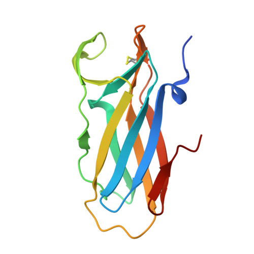
6DPS

Crystal Structure of Neisseria meningitidis DsbD n-terminal domain in the oxidised form


Experimental Data Snapshot

  • Method: X-RAY DIFFRACTION
  • Resolution: 2.56 Å
  • R-Value Free: 0.235 
  • R-Value Work: 0.178 
  • R-Value Observed: 0.181 

wwPDB Validation   3D Report Full Report


This is version 1.3 of the entry. See complete history


Literature

Structural and biochemical insights into the disulfide reductase mechanism of DsbD, an essential enzyme for neisserial pathogens.

Smith, R.P.Mohanty, B.Mowlaboccus, S.Paxman, J.J.Williams, M.L.Headey, S.J.Wang, G.Subedi, P.Doak, B.C.Kahler, C.M.Scanlon, M.J.Heras, B.

(2018) J Biol Chem 293: 16559-16571

  • DOI: https://doi.org/10.1074/jbc.RA118.004847
  • Primary Citation of Related Structures:  
    6DNL, 6DNU, 6DNV, 6DPS

  • PubMed Abstract: 

    The worldwide incidence of neisserial infections, particularly gonococcal infections, is increasingly associated with antibiotic-resistant strains. In particular, extensively drug-resistant Neisseria gonorrhoeae strains that are resistant to third-generation cephalosporins are a major public health concern. There is a pressing clinical need to identify new targets for the development of antibiotics effective against Neisseria -specific processes. In this study, we report that the bacterial disulfide reductase DsbD is highly prevalent and conserved among Neisseria spp. and that this enzyme is essential for survival of N. gonorrhoeae DsbD is a membrane-bound protein that consists of two periplasmic domains, n-DsbD and c-DsbD, which flank the transmembrane domain t-DsbD. In this work, we show that the two functionally essential periplasmic domains of Neisseria DsbD catalyze electron transfer reactions through unidirectional interdomain interactions, from reduced c-DsbD to oxidized n-DsbD, and that this process is not dictated by their redox potentials. Structural characterization of the Neisseria n- and c-DsbD domains in both redox states provides evidence that steric hindrance reduces interactions between the two periplasmic domains when n-DsbD is reduced, thereby preventing a futile redox cycle. Finally, we propose a conserved mechanism of electron transfer for DsbD and define the residues involved in domain-domain recognition. Inhibitors of the interaction of the two DsbD domains have the potential to be developed as anti-neisserial agents.


  • Organizational Affiliation

    From the Department of Biochemistry and Genetics, La Trobe Institute for Molecular Science, La Trobe University, Melbourne 3086, Victoria, Australia.


Macromolecules
Find similar proteins by:  (by identity cutoff)  |  3D Structure
Entity ID: 1
MoleculeChains Sequence LengthOrganismDetailsImage
Thiol:disulfide interchange protein DsbD
A, B, C, D, E
A, B, C, D, E, F
126Neisseria meningitidis alpha14Mutation(s): 1 
Gene Names: dsbDNMO_1340
EC: 1.8.1.8
UniProt
Find proteins for C6S7X6 (Neisseria meningitidis (strain alpha14))
Explore C6S7X6 
Go to UniProtKB:  C6S7X6
Entity Groups  
Sequence Clusters30% Identity50% Identity70% Identity90% Identity95% Identity100% Identity
UniProt GroupC6S7X6
Sequence Annotations
Expand
  • Reference Sequence
Small Molecules
Ligands 1 Unique
IDChains Name / Formula / InChI Key2D Diagram3D Interactions
ZN
Query on ZN

Download Ideal Coordinates CCD File 
AA [auth D]
BA [auth D]
CA [auth D]
DA [auth D]
EA [auth E]
AA [auth D],
BA [auth D],
CA [auth D],
DA [auth D],
EA [auth E],
FA [auth E],
G [auth A],
GA [auth F],
H [auth A],
HA [auth F],
I [auth A],
IA [auth F],
J [auth B],
JA [auth F],
K [auth B],
KA [auth F],
L [auth B],
LA [auth F],
M [auth B],
N [auth B],
O [auth B],
P [auth B],
Q [auth B],
R [auth C],
S [auth C],
T [auth C],
U [auth C],
V [auth C],
W [auth C],
X [auth D],
Y [auth D],
Z [auth D]
ZINC ION
Zn
PTFCDOFLOPIGGS-UHFFFAOYSA-N
Experimental Data & Validation

Experimental Data

  • Method: X-RAY DIFFRACTION
  • Resolution: 2.56 Å
  • R-Value Free: 0.235 
  • R-Value Work: 0.178 
  • R-Value Observed: 0.181 
  • Space Group: P 21 3
Unit Cell:
Length ( Å )Angle ( ˚ )
a = 147.643α = 90
b = 147.643β = 90
c = 147.643γ = 90
Software Package:
Software NamePurpose
HKL-2000data scaling
SOLVEphasing
PHENIXrefinement
PDB_EXTRACTdata extraction
HKL-2000data reduction

Structure Validation

View Full Validation Report


Entry History 

Deposition Data

Revision History  (Full details and data files)

  • Version 1.0: 2018-09-12
    Type: Initial release
  • Version 1.1: 2018-09-19
    Changes: Data collection, Database references
  • Version 1.2: 2018-11-07
    Changes: Data collection, Database references
  • Version 1.3: 2024-11-20
    Changes: Data collection, Database references, Derived calculations, Structure summary