


Structure of a heterodimer of neuronal cell surface proteins
Ranaivoson, F.M., Turk, L.S., Ozkan, E., Montelione, G.T., Comoletti, D.(2019) Structure
Experimental Data Snapshot
Starting Model: experimental
View more details
(2019) Structure
Entity ID: 1 | |||||
|---|---|---|---|---|---|
| Molecule | Chains | Sequence Length | Organism | Details | Image |
| IgLON family member 5 | 303 | Homo sapiens | Mutation(s): 0 Gene Names: IGLON5 | 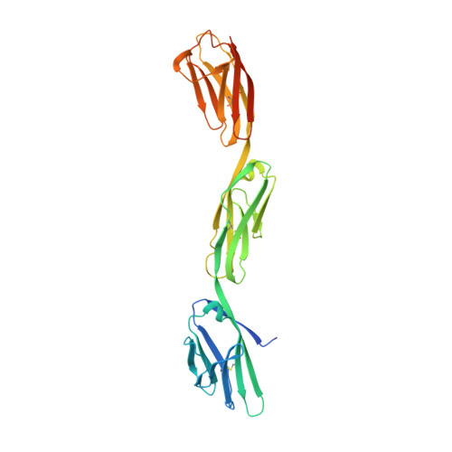 | |
UniProt & NIH Common Fund Data Resources | |||||
PHAROS: A6NGN9 GTEx: ENSG00000142549 | |||||
Entity Groups | |||||
| Sequence Clusters | 30% Identity50% Identity70% Identity90% Identity95% Identity100% Identity | ||||
| UniProt Group | A6NGN9 | ||||
Glycosylation | |||||
| Glycosylation Sites: 5 | Go to GlyGen: A6NGN9-1 | ||||
Sequence AnnotationsExpand | |||||
Reference Sequence | |||||
Entity ID: 2 | |||||
|---|---|---|---|---|---|
| Molecule | Chains | Sequence Length | Organism | Details | Image |
| Neuronal growth regulator 1 | 304 | Homo sapiens | Mutation(s): 0 Gene Names: NEGR1, IGLON4, UNQ2433/PRO4993 | 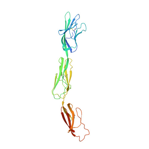 | |
UniProt & NIH Common Fund Data Resources | |||||
PHAROS: Q7Z3B1 GTEx: ENSG00000172260 | |||||
Entity Groups | |||||
| Sequence Clusters | 30% Identity50% Identity70% Identity90% Identity95% Identity100% Identity | ||||
| UniProt Group | Q7Z3B1 | ||||
Glycosylation | |||||
| Glycosylation Sites: 3 | Go to GlyGen: Q7Z3B1-1 | ||||
Sequence AnnotationsExpand | |||||
Reference Sequence | |||||
Entity ID: 3 | |||||
|---|---|---|---|---|---|
| Molecule | Chains | Length | 2D Diagram | Glycosylation | D Interactions |
| alpha-D-mannopyranose-(1-3)-beta-D-mannopyranose-(1-4)-2-acetamido-2-deoxy-beta-D-glucopyranose-(1-4)-2-acetamido-2-deoxy-beta-D-glucopyranose | E | 4 |  | N-Glycosylation | |
Glycosylation Resources | |||||
GlyTouCan: G81315DD GlyCosmos: G81315DD GlyGen: G81315DD | |||||
| Ligands 1 Unique | |||||
|---|---|---|---|---|---|
| ID | Chains | Name / Formula / InChI Key | 2D Diagram | 3D Interactions | |
| NAG Download:Ideal Coordinates CCD File | F [auth A] G [auth A] H [auth A] I [auth A] J [auth B] | 2-acetamido-2-deoxy-beta-D-glucopyranose C8 H15 N O6 OVRNDRQMDRJTHS-FMDGEEDCSA-N |  | ||
| Length ( Å ) | Angle ( ˚ ) |
|---|---|
| a = 120.341 | α = 90 |
| b = 305.799 | β = 100.78 |
| c = 60.029 | γ = 90 |
| Software Name | Purpose |
|---|---|
| PHENIX | refinement |
| XDS | data reduction |
| XDS | data scaling |
| PHASER | phasing |
| Funding Organization | Location | Grant Number |
|---|---|---|
| National Science Foundation (NSF, United States) | United States | MCB-1450895 |


