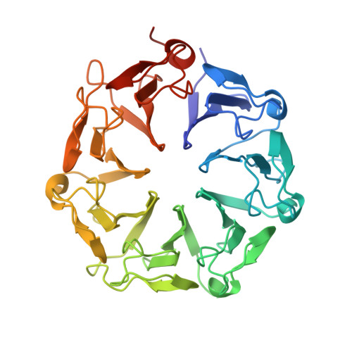
6DD7

Crystal structure of plant UVB photoreceptor UVR8 from in situ serial Laue diffraction


Experimental Data Snapshot

  • Method: X-RAY DIFFRACTION
  • Resolution: 2.00 Å
  • R-Value Free: 0.328 
  • R-Value Work: 0.254 
  • R-Value Observed: 0.257 

wwPDB Validation   3D Report Full Report


This is version 1.3 of the entry. See complete history


Literature

Crystal-on-crystal chips for in situ serial diffraction at room temperature.

Ren, Z.Ayhan, M.Bandara, S.Bowatte, K.Kumarapperuma, I.Gunawardana, S.Shin, H.Wang, C.Zeng, X.Yang, X.

(2018) Lab Chip 18: 2246-2256

  • DOI: https://doi.org/10.1039/c8lc00489g
  • Primary Citation of Related Structures:  
    6DD6, 6DD7

  • PubMed Abstract: 

    Recent developments in serial crystallography at X-ray free electron lasers (XFELs) and synchrotrons have been driven by two scientific goals in structural biology - first, static structure determination from nano or microcrystals of membrane proteins and large complexes that are difficult for conventional cryocrystallography, and second, direct observations of transient structural species in biochemical reactions at near atomic resolution. Since room-temperature diffraction experiments naturally demand a large quantity of purified protein, sample economy is critically important for all steps of serial crystallography from crystallization, crystal delivery to data collection. Here we report the development and applications of "crystal-on-crystal" devices to facilitate large-scale in situ serial diffraction experiments on protein crystals of all sizes - large, small, or microscopic. We show that the monocrystalline quartz as a substrate material prevents vapor loss during crystallization and significantly reduces background X-ray scattering. These devices can be readily adopted at XFEL and synchrotron beamlines, which enable efficient delivery of hundreds to millions of crystals to the X-ray beam, with an overall protein consumption per dataset comparable to that of cryocrystallography.


  • Organizational Affiliation

    Department of Chemistry, The University of Illinois at Chicago, Chicago, IL 60607, USA. zren@uic.edu xiaojing@uic.edu.


Macromolecules
Find similar proteins by:  (by identity cutoff)  |  3D Structure
Entity ID: 1
MoleculeChains Sequence LengthOrganismDetailsImage
Ultraviolet-B receptor UVR8
A, B, C, D
377Arabidopsis thalianaMutation(s): 0 
Gene Names: UVR8At5g63860MGI19.7
UniProt
Find proteins for Q9FN03 (Arabidopsis thaliana)
Explore Q9FN03 
Go to UniProtKB:  Q9FN03
Entity Groups  
Sequence Clusters30% Identity50% Identity70% Identity90% Identity95% Identity100% Identity
UniProt GroupQ9FN03
Sequence Annotations
Expand
  • Reference Sequence
Experimental Data & Validation

Experimental Data

  • Method: X-RAY DIFFRACTION
  • Resolution: 2.00 Å
  • R-Value Free: 0.328 
  • R-Value Work: 0.254 
  • R-Value Observed: 0.257 
  • Space Group: C 1 2 1
Unit Cell:
Length ( Å )Angle ( ˚ )
a = 111.2α = 90
b = 80.3β = 94.8
c = 190γ = 90
Software Package:
Software NamePurpose
PHENIXrefinement
Precognitiondata reduction
Epinormdata reduction

Structure Validation

View Full Validation Report


Entry History & Funding Information

Deposition Data

  • Released Date: 2018-07-18 
  • Deposition Author(s): Ren, Z.

Funding OrganizationLocationGrant Number
National Institutes of Health/National Eye Institute (NIH/NEI)United StatesR01EY024363

Revision History  (Full details and data files)

  • Version 1.0: 2018-07-18
    Type: Initial release
  • Version 1.1: 2018-08-08
    Changes: Data collection, Database references
  • Version 1.2: 2019-12-11
    Changes: Author supporting evidence
  • Version 1.3: 2023-10-11
    Changes: Data collection, Database references, Refinement description