6DB9 | pdb_00006db9

Structural basis for promiscuous binding and activation of fluorogenic dyes by DIR2s RNA aptamer


Experimental Data Snapshot

  • Method: X-RAY DIFFRACTION
  • Resolution: 2.02 Å
  • R-Value Free: 
    0.257 (Depositor), 0.259 (DCC) 
  • R-Value Work: 
    0.230 (Depositor), 0.233 (DCC) 
  • R-Value Observed: 
    0.231 (Depositor) 

Starting Model: experimental
View more details

wwPDB Validation 3D Report Full Report

Validation slider image for 6DB9

This is version 1.3 of the entry. See complete history

Literature

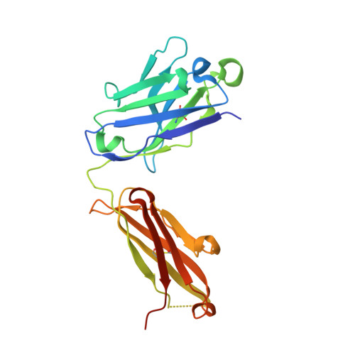
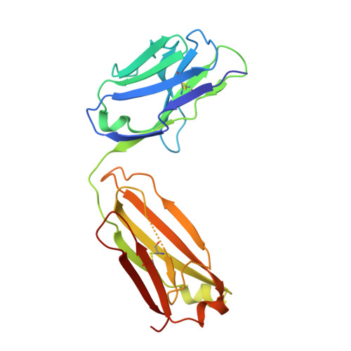
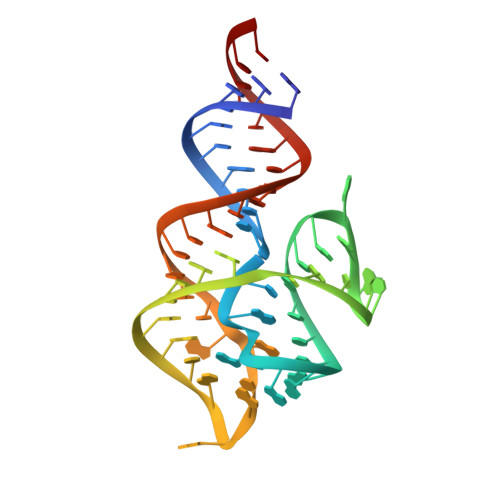
Structural basis for activation of fluorogenic dyes by an RNA aptamer lacking a G-quadruplex motif.

Shelke, S.A.Shao, Y.Laski, A.Koirala, D.Weissman, B.P.Fuller, J.R.Tan, X.Constantin, T.P.Waggoner, A.S.Bruchez, M.P.Armitage, B.A.Piccirilli, J.A.

(2018) Nat Commun 9: 4542-4542

  • DOI: https://doi.org/10.1038/s41467-018-06942-3
  • Primary Citation Related Structures: 
    6DB8, 6DB9

  • PubMed Abstract: 

    The DIR2s RNA aptamer, a second-generation, in-vitro selected binder to dimethylindole red (DIR), activates the fluorescence of cyanine dyes, DIR and oxazole thiazole blue (OTB), allowing detection of two well-resolved emission colors. Using Fab BL3-6 and its cognate hairpin as a crystallization module, we solved the crystal structures of both the apo and OTB-SO 3 bound forms of DIR2s at 2.0 Å and 1.8 Å resolution, respectively. DIR2s adopts a compact, tuning fork-like architecture comprised of a helix and two short stem-loops oriented in parallel to create the ligand binding site through tertiary interactions. The OTB-SO 3 fluorophore binds in a planar conformation to a claw-like structure formed by a purine base-triple, which provides a stacking platform for OTB-SO 3, and an unpaired nucleotide, which partially caps the binding site from the top. The absence of a G-quartet or base tetrad makes the DIR2s aptamer unique among fluorogenic RNAs with known 3D structure.


  • Organizational Affiliation
    • Department of Biochemistry and Molecular Biology, The University of Chicago, Chicago, IL, 60637, USA.

Macromolecule Content 

  • Total Structure Weight: 67.38 kDa 
  • Atom Count: 4,682 
  • Modeled Residue Count: 488 
  • Deposited Residue Count: 506 
  • Unique protein chains: 2
  • Unique nucleic acid chains: 1

Macromolecules


Find similar proteins by:|  3D Structure
Entity ID: 2
MoleculeChains  Sequence LengthOrganismDetailsImage
Fab-Heavy-ChainB [auth H]232synthetic constructMutation(s): 0 
Find similar proteins by:|  3D Structure
Entity ID: 3
MoleculeChains  Sequence LengthOrganismDetailsImage
Fab-Light-ChainC [auth L]214synthetic constructMutation(s): 0 
Find similar nucleic acids by:  (by identity cutoff) 
Entity ID: 1
MoleculeChains LengthOrganismImage
RNA (60-MER)A [auth R]60synthetic construct
Sequence Annotations
Expand
Reference Sequence

Experimental Data & Validation

Experimental Data

  • Method: X-RAY DIFFRACTION
  • Resolution: 2.02 Å
  • R-Value Free:  0.257 (Depositor), 0.259 (DCC) 
  • R-Value Work:  0.230 (Depositor), 0.233 (DCC) 
  • R-Value Observed: 0.231 (Depositor) 
Space Group: P 21 21 21
Unit Cell:
Length ( Å )Angle ( ˚ )
a = 83.78α = 90
b = 83.805β = 90
c = 109.926γ = 90
Software Package:
Software NamePurpose
PHENIXrefinement
PHENIXphasing
Cootmodel building

Structure Validation

View Full Validation Report


Entry History 

& Funding Information

Deposition Data


Funding OrganizationLocationGrant Number
National Institutes of Health/National Human Genome Research Institute (NIH/NHGRI)United StatesR01AI081987

Revision History  (Full details and data files)

  • Version 1.0: 2018-11-14
    Type: Initial release
  • Version 1.1: 2019-12-18
    Changes: Author supporting evidence
  • Version 1.2: 2023-10-04
    Changes: Data collection, Database references, Derived calculations, Refinement description
  • Version 1.3: 2024-10-23
    Changes: Structure summary